Anatomical regurgitant orifice area in aortic regurgitation: a quantitative key to severity assessment?
European Heart Journal


Abstract
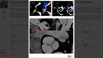
Aortic regurgitation (AR) is challenging to assess. Transthoracic echocardiography (TTE) is the primary tool for diagnosing AR, requiring a multiparametric approach with both qualitative and quantitative data. Severe AR often causes left ventricular (LV) dilation due to volume overload, making LV size and ejection fraction key indicators of severity. Cardiac CT, though not widely incorporated in current guidelines, may offer value by measuring the anatomical regurgitant orifice (ARO). However, the clinical significance of ARO remains uncertain due to limited research.
The aim of this study was to assess if ARO, measured via cardiac CT, correlates with LV volume overload in severe AR patients. A secondary goal was to identify echocardiographic parameters that predict ARO size. We retrospectively enrolled 37 patients with severe AR by TTE, all of whom underwent cardiac CT within 12 months. Two blinded cardiologists measured the ARO area planimetry. The average ARO area in diastole was 0.33±0.03 cm². A strong correlation was found between ARO and LV telediastolic volume (LVTDV) measured by CT (r=0.7, p<0.05) and TTE (r=0.65, p<0.05). Moderate correlations were observed with LV telesystolic volume (LVTSV): r=0.45 in CT and r=0.5 in TTE (p<0.05). ARO also showed a moderate to strong correlation with LV telediastolic diameter (LVTDD) and a weak correlation with LV telesystolic diameter (LVTSD): r=0.6 and r=0.3 respectively (p<0.01).
These results suggest that ARO correlates most strongly with LV dilation in diastole, reflecting AR hemodynamic impact. In the regression analysis, vena contracta width was the strongest predictor of ARO (R²=0.57, p<0.01), followed by jet/LVOT ratio (R²=0.47, p<0.01) and peak end-diastolic velocity in descending aorta (R²=0.45, p<0.01). These findings suggest that ARO is influenced by both anatomical and hemodynamic factors, which affect AR severity. This study confirms that ARO by CT is a valuable marker for assessing AR severity and closely correlates with LV volume overload. Additionally, the weaker correlation with telesystolic measurements can be explained by the fact that not all patients were at the late stages of AR, where systolic dilation and reduced LV ejection fraction (LVEF) develop. This is relevant, as LVEF and LVTSD are key criteria for surgical intervention in current guidelines.
In conclusion, the ARO area is a quantitative parameter that can be especially useful in cases where multimodal imaging results for AR are inconclusive. The correlation of ARO with LV dilation reflects a clear pathophysiological relationship, underscoring its clinical and prognostic significance and its utility in risk stratification. Furthermore, the correlation between ARO and echocardiographic indices of AR severity reinforces its value as a robust diagnostic tool, bridging both anatomical and hemodynamic assessments. TTE and CT parameters LV volumes and ARO area correlation
Contributors



You may be interested in