Liver native T1 mapping for predicting cardiovascular outcomes across stages of heart failure
European Heart Journal


Abstract
Patients who progressive stages of heart failure (HF) may have a risk of developing liver inflammation and fibrosis. Magnetic resonance imaging (MRI)-based liver T1 mapping quantifies the underlying inflammatory and fibrotic status.
We aimed to explore the associations of liver T1mapping with risk of cardiovascular events across heart failure (HF) stages.
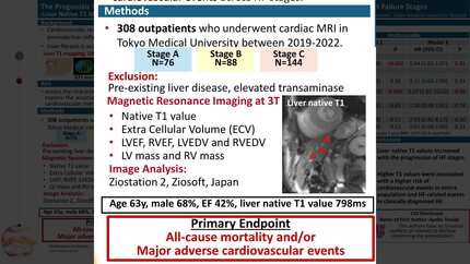
This retrospective study included 308 individuals who underwent cardiac MRI and were classified into HF stages according to American College of Cardiology/American Heart Association guidelines. Those with pre-existing liver disease were excluded. Primary outcome was a composite of all-cause mortality and/or major adverse cardiovascular events.
Mean age was 63±15years, 68% male, 47% had HF stage C, and mean left ventricular ejection fraction (EF) was 42±18%. Mean liver native T1 value was 798±131ms and was higher as HF stages progressed (P<0.01). Over a median follow-up of 18.2 months, 9.4% experienced the primary outcome. After covariate adjustment (i.e., B-type natriuretic peptide, and left and right ventricular EF), higher liver native T1 values were associated with an increased risk of the primary outcome (adjusted-HR: 1.04; 95%CI:1.01-1.07; P=0.01), which was consistent across HF stages (P-for-interaction>0.10). Among patients with HF stage C, higher liver native T1 values were associated with an increased risk of all-cause mortality and/or HF hospitalization (adjusted-HR: 1.01; 95% CI: 1.00-1.01; P<0.001; P-for-interaction<0.001).
Liver native T1 values increased with the progression of HF stages. Higher T1 values predicted the risk of cardiovascular events in entire population and HF-related events in clinically diagnosed HF.
Contributors


You may be interested in