Insight into left ventricular diastolic compensation in hypertrophic cardiomyopathy with left ventricular outflow obstruction: a 4D flow CMR study
European Heart Journal


Abstract
Presence of echo-assessed left ventricular outflow tract obstruction (LVOTO) increases risks of heart failure and death in hypertrophic cardiomyopathy (HCM). Four-dimensional (4D) flow cardiac magnetic resonance (CMR) which enables whole-volume intracardiac hemodynamic characterization, may offer complementary mechanistic insights.
Compare 4D flow CMR-assessed intracardiac flow and kinetic energy (KE) in distinct HCM phenotypes.
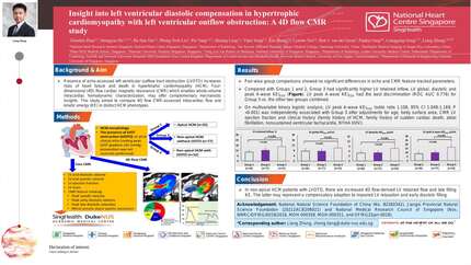
We retrospectively analyzed 165 clinically diagnosed HCM patients at a single institution who had undergone cine and 4D flow CMR. Based on HCM morphology and report of the presence of LVOTO on prior clinical echo (resting peak LVOT gradient ≥30 mmHg; provocation was not routinely performed), we stratified subjects into three groups: Group 1, apical HCM (n=36); Group 2, non-apical HCM without LVOTO (n=77); Group 3, non-apical HCM with LVOTO (n=52). We extracted values of early (E) and late (A) diastolic mitral filling velocities, early (e’) and late diastolic mitral annular velocities, and E/e’ from echo reports. We performed all CMR analyses in a core lab, including: (1) 4D flow-derived flow components (direct flow, retained inflow, delayed ejection flow, and residual volume) and phasic KE normalized to left ventricular (LV) end-diastolic volume (KEiEDV) (global, peak systolic, systolic, diastolic, peak E-wave and peak A-wave); and (2) averaged values of peak systolic velocities, peak early diastolic velocities, late diastolic velocities, and mitral annular plane systolic excursions extracted from velocity or displacement curves of mitral annular motion in the two- and four-chamber views obtained using an in-house CMR feature tracking algorithm.
Except for Group 3 subjects having a smaller body surface area, all three groups had similar age and proportions of subjects with family history of HCM, family history of sudden cardiac death, atrial fibrillation, and nonsustained ventricular tachycardia. Pair-wise group comparisons showed no significant differences in echo and CMR feature-tracked parameters. Compared with Groups 1 and 2, Group 3 had significantly higher LV retained inflow, LV global, diastolic and peak A-wave KEiEDV (Figure). LV peak A-wave KEiEDV had the best discrimination (ROC AUC 0.776) for Group 3 vs. the other two groups combined. On multivariable binary logistic analysis, LV peak A-wave KEiEDV (odds ratio 1.108, 95% CI 1.049-1.169; P <0.001) was independently associated with Group 3 after adjustments for age, body surface area, CMR LV ejection fraction and clinical history (family history of HCM, family history of sudden cardiac death, atrial fibrillation, nonsustained ventricular tachycardia, NYHA III/IV).
In non-apical HCM patients with LVOTO, there are increased 4D flow-derived LV retained flow and late filling KE. The latter may represent a compensatory adaption to impaired LV relaxation and early diastolic filling.



You may be interested in