Brain amyloid accumulation in patients with transthyretin amyloid cardiomyopathy: a pilot study
European Heart Journal


Abstract
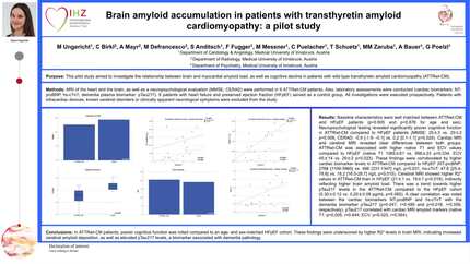
Wild-type transthyretin amyloid cardiomyopathy (ATTRwt-CM) and dementia are both age-related protein-misfolding disorders. However, a causal relationship in human has not yet been established. The aim of this pilot study was to investigate the relationship between brain and myocardial amyloid accumulation, as well as cognitive decline in patients with ATTRwt-CM.
Magnetic resonance imaging (MRI) of the heart and the brain, as well as a comprehensive neuropsychological evaluation (CERAD-Plus, MMSE) were performed in 9 ATTRwt-CM patients. Also, laboratory assessments were conducted (cardiac biomarkers: NT-proBNP, hs-cTnT; dementia plasma biomarker: pTau217). 9 patients with heart failure and preserved ejection fraction (HFpEF) served as a control group. All investigations were executed prospectively, with patients recruited consecutively at the Medical University Innsbruck. Patients with intracardiac devices, known cerebral disorders, or clinically apparent neurological symptoms were excluded from the study.
Baseline characteristics were well matched between ATTRwt-CM and HFpEF (p=0.805 and p=0.876 for age and sex). Neuropsychological testing revealed significantly poorer cognitive function in ATTRwt-CM compared to HFpEF patients (MMSE: 25 [22-28] vs. 29 [27.5-30] p=0.005, CERAD-Plus: -0.9 [-2.3- -0.1] vs. 0.2 [-0.1-1.1] p=0.014). Cardiac MRI and cerebral MRI revealed clear differences between both groups. ATTRwt-CM was associated with higher native T1 and ECV values compared to HFpEF (native T1 1065±61 vs. 998±23 p=0.034, ECV 45±14 vs. 29±2 p=0.023). These findings were corroborated by higher cardiac biomarker levels in ATTRwt-CM compared to HFpEF (NT-proBNP: 2585 [1146-3819] vs. 498 [331-1347] ng/L p=0.027, hs-cTnT: 47.1 [27.3-78.6] vs. 18.2 [16.5-28.7] ng/L p=0.011). Cerebral MRI showed higher R2* values in ATTRwt-CM than in HFpEF (21±1 vs. 19±1 p=0.019), indirectly reflecting higher brain amyloid load. No correlations were found between cardiac (native T1, ECV) and cerebral amyloid markers (R2*) (p>0.05 for all comparisons). There was a trend towards higher pTau217 levels in the ATTRwt-CM compared to the HFpEF cohort (0.30±0.12 vs. 0.20±0.09 pg/mL p=0.065). A clear correlation was noted between the cardiac biomarkers NT-proBNP and hs-cTnT with the dementia biomarker pTau217 (p=0.040, r=0.487 and p=0.016, r=0.559, respectively). pTau217 correlated with cardiac MRI amyloid markers (native T1: p=0.005, r=0.644; ECV: p=0.023, r=0.564).
In ATTRwt-CM patients, poorer cognitive function was noted compared to an age- and sex-matched HFpEF cohort. These findings were underscored by higher R2* levels in brain MRI, indicating increased cerebral amyloid deposition, as well as elevated pTau217 levels, a biomarker associated with dementia pathology. No direct correlations were found between the extent of brain (R2* in brain MRI) and myocardial amyloid burden (native T1, ECV in cardiac MRI).
Contributors


C Birkl
Author
A Mayr
Author
M Defrancesco
Author
S Anditsch
Author
F Fugger
Author
M Messner
Author
C Puelacher
Author
T Schuetz
Author
M M Zaruba
Author
A Bauer
Author
G Poelzl
Author
You may be interested in