Comprehensive cardiovascular magnetic resonance for evaluating structural myocardial changes in patients with hypertrophic obstructive cardiomyopathy treated with the first myosin inhibitor
European Heart Journal


Abstract
Mavacamten is the first myosin inhibitor targeting hypercontractility in hypertrophic obstructive cardiomyopathy (HOCM). Despite known functional improvements, results on its effects on myocardial structure based on cardiovascular magnetic resonance (CMR) are sparse. This study aims to evaluate the one-year structural effects of mavacamten assessed by comprehensive CMR examinations.
In this ongoing prospective study all included HOCM patients undergo comprehensive contrast-enhanced 1.5 T-CMR at baseline and one-year follow-up. Following current guidelines recommendations, patients either remain on baseline negative inotropic therapy or receive additional treatment with the first myosin inhibitor mavacamten. Key parameters, including left and right ventricular ejection fraction (LVEF/RVEF), cardiac index (CI), LV mass, global longitudinal strain (GLS), late gadolinium enhancement (LGE, %), native T1 mapping, and extracellular volume (ECV) are compared between both groups at baseline and follow-up.
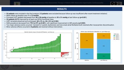
To date, 19 patients have been enrolled (mean age 59.0 ± 12.0 years, 73.6% male). Mavacamten therapy was initiated in 11/19 patients (58%). Compared to the control group, patients treated with mavacamten showed a significant greater reduction in LV mass index (-28.0 g/m² [95% CI -43.4 to -12.6] vs. +3.4 g/m² [95% CI -2.4 to +9.1], p=0.004) and left atrial (LA) volume index (-15.5 mL/m² [95% CI -2.7 to -28.3] vs. +4.2 mL/m² [95% CI -6.4 to +14.8], p=0.008). Moreover, in the mavacamten group, there was a significant reduction in LVEF (79.3 ± 4.8% to 70.5 ± 8.8%, p=0.029), RVEF (48.3 ± 7.4% to 42.5 ± 6.0%, p=0.011), and CI (3.0 ± 0.8 to 2.5 ± 0.7 L/min/m², p=0.020) at the time of follow up. In contrast, the control group showed stable LVEF and RVEF and a significant increase in CI (LVEF: 76.1 ± 11.2% to 76.3 ± 12%, p=0.724; RVEF: 49.4 ± 6.8% to 52.1 ± 8.8%, p=0.192; CI: 2.0 ± 0.5 to 2.4 ± 0.6 L/min/m², p=0.012). GLS of both LV and RV showed no significant changes within or between groups (GLS LV: +3.34 [95% CI +10.0 to -3.3] vs. -0.8 [95% CI -2.4 to -0.8] %, p=0.457; GLS RV: +5.3 [95% CI +10.2 to +0.4] vs. +1.6 [95% CI +7 to -4.1] %, p=0.409).
Regarding myocardial fibrosis, the mavacamten group showed a significant reduction in global myocardial native T1 (1040.1 ± 23.2 vs. 1001.9 ± 36.0, p=0.016) and LGE extent (9.1 ± 6.0% to 8.8 ± 6.0%, p=0.017) while ECV remained unchanged (25.0 ± 2.9 vs. 28.2 ± 5.0, p=0.151). In contrast, all parameters remained unaltered in the control group.
Myosin inhibitor therapy favourably affects structural remodelling by reducing LV mass and left LA size. While LVEF and CI decrease, the preservation of GLS suggests maintained regional systolic performance during myosin inhibition. Additionally, our findings indicate the potential of a slight reduction in fibrosis, though a larger cohort is required to validate these effects.


You may be interested in