A validation study of novel full free-breathing cardiac magnetic resonance comparing to traditional breath-holding method: correlation of cardiac magnetic resonance-derived multi parameters
European Heart Journal


Abstract
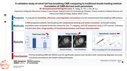
Conventional cardiac magnetic resonance (CMR) imaging typically requires patients to hold their breath, with nearly 50% of the total scan time spent resting between breath-holds. This practice limits the widespread adoption of CMR, especially in settings where breath-holding is difficult for patients. Furthermore, suboptimal image quality often results from poor breath-holding, which can compromise diagnostic accuracy. The study has advanced a novel full free-breathing CMR techniques, incorporating compressed sensing and motion correction (Moco) technologies, which can significantly shorten the scanning time.
The aim of this study is to evaluate whether novel full free-breathing CMR can replace traditional breath-holding method while maintaining diagnostic quality.
Patients who underwent full free-breathing CMR examinations between June 1 and 30, 2024, were prospectively included in the analysis. Key parameters, including image acquisition time and left ventricular function measurements, were compared with those obtained from traditional breath-holding CMR. Additionally, data from full free-breathing and breath-holding T1 mapping and late gadolinium enhancement (LGE) scans conducted between November 15 and December 14 were analysed. Group comparisons were made using the Wilcoxon signed-rank test or paired t-test. Consistency testing was performed using Kappa, Bland-Altman, and linear regression analyses.
A total of 282 patients (median age, 53 years [38, 63]; range, 10-82 years; 154 men) were evaluated. The mean acquisition time for full free-breathing CMR was 22.6±3.7 minutes. Compared with traditional breath-holding CMR, measurements of the end-diastolic volume (EDV), end-systolic volume (ESV), EDV index, and ESV index obtained using free-breathing CMR were slightly higher (all P<0.05), while left ventricular ejection fraction (LVEF) and left ventricular mass (LVM) were slightly lower (both P<0.05) . However, there was good consistency and correlation between the two methods (r values: 0.82–0.98) (Figure 1). Native T1 and LGE values were also highly correlated (r values: 0.83–0.98) (Figure 2).
Full free-breathing CMR is feasible and effective in clinical practice. It significantly shortens examination time and can replace conventional breath-holding CMR without compromising diagnostic accuracy.

You may be interested in