Combination of pre-operative computed tomography-derived longitudinal strain and extracellular volume fraction is a useful prognostic marker after transcatheter aortic valve replacement
European Heart Journal


Abstract
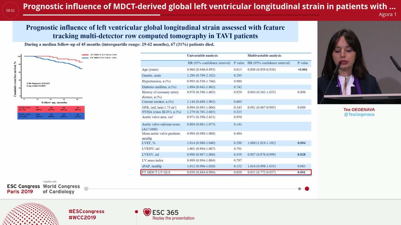
Using state-of-the-art image analysis software, strain and Extracellular volume fraction (ECV) analysis for left ventricular (LV) myocardium (LVM) can also be performed using cardiac computed tomography (CT) images.
This study aims to evaluate the utility of myocardial strain and ECV analysis on CT to predict the prognosis of patients who underwent transcatheter aortic valve replacement (TAVR).
We included 126 patients with severe aortic valve stenosis (AS) who underwent preoperative cardiac CT. The major adverse cardiac events (MACE)s were defined as the composite of cardiovascular death and hospitalization due to heart failure (HF) or fatal arrhythmia during the follow-up. During the follow-up period (651±383 days), twenty-four patients (19%) had MACEs. Receiver operating characteristic (ROC) curve analysis was performed to evaluate the diagnostic performance of ECV and GLS of LVM on CT for predicting prognosis. Optimal cut-off values of ECV and GLS of LVM were 32.43% and -9.98% based on the ROC analysis (P= 0.0247 and 0.0007) (Figures A and B), and all patients were subsequently categorized into three groups: the patients with ECV ≥ 32.43% and GLS ≥ -9.98% (n=14), those with either ECV ≥ 32.43% or GLS ≥ -9.98% (n=40), and those with neither ECV ≥ 32.43% nor GLS ≥ -9.98% (n=72). Among the three groups, patients with both pre-operative ECV ≥ 32.43% and GLS > -9.98% demonstrated the worst prognosis, with a significantly lower survival rate according to the Kaplan-Meier survival curves and log-rank test (p < 0.05) (Figure C).
In the Cox proportional hazard model, patients with both pre-operative ECV≥ 32.43% and GLS≤ -9.98% had a significantly higher risk of cardiovascular events (hazard ratio (HR), 5.10; 95% CI, 2.06-11.65; P =0.0009).
Our findings indicate that the combination of ECV and GLS was a sensitive predictor of prognosis after TAVR. TAVI CT strain figure
Contributors

H Takaoka
Author
S Aoki
Author
Y Noguchi
Author
Y Nishikawa
Author
K Yoshida
Author
K Suzuki
Author
S Yashima
Author
M Kinoshita
Author
H Sasaki
Author
N Suzuki-Eguchi
Author
S Okita
Author
H Kitahara
Author
Y Kobayashi
Author
You may be interested in