Comparison of false positive rates and radiation exposure in dual-source vs. photon-counting cardiac CT for detecting severe coronary stenosis: a retrospective cohort study
European Heart Journal


Abstract
Cardiac computed tomography angiography (CCTA) is a key non-invasive tool for assessing coronary artery disease (CAD). The 2024 European Society of Cardiology (ESC) guidelines highlight its growing role, particularly for patients with low to moderate pre-test probability. However, its high sensitivity can lead to false positive (FP) findings, resulting in unnecessary invasive coronary angiographies (ICA). Photon-counting computed tomography (PC-CT) offers potential benefits over standard dual-source CT (DS-CT), including improved resolution, enhanced contrast, and lower radiation exposure. This study compares DS-CT and PC-CT in terms of FP rates and radiation dose.
This study aims to compare false positive rates and radiation exposure between DS-CT and PC-CT in detecting severe coronary stenosis. By assessing diagnostic accuracy and radiation dose, we seek to determine whether PC-CT offers advantages over DS-CT in clinical practice.
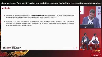
This retrospective cohort study included consecutive patients between 2019 and 2024 who underwent CCTA for suspected CAD with a positive finding for severe coronary stenosis. Patients who had ICA at the same institution within three months of CCTA were included. A positive CCTA result was defined as 70–99% stenosis in a major coronary artery or bypass graft, left main artery stenosis of 50% or more, three-vessel disease with stenosis of 70% or more, or chronic total occlusion. Significant stenosis on ICA was defined as at least 50% stenosis in the left main coronary artery, at least 70% stenosis in a major coronary artery or bypass graft, or stenosis between 30–70% with physiological significance (FFR ≤0.8 or iFR ≤0.89). False positive rates between DS-CT and PC-CT were compared using chi-square tests, while radiation exposure was assessed by dose-length product (DLP, mGy×cm). A binomial logistic regression model evaluated differences between imaging techniques. Baseline characteristics, including BMI and calcium score, were similar between groups.
261 patients met the inclusion criteria, of which 141 (54.0%) underwent DS-CT and 120 (46.0%) underwent PC-CT. The FP rate was higher in DS-CT (40/141, 15.3%) compared to PC-CT (20/120, 7.7%) (p=0.025). Radiation exposure, measured as DLP, was significantly lower in PC-CT compared to DS-CT. The mean DLP for PC-CT was 271 mGy×cm (range: 50–1386), whereas for DS-CT, the mean DLP was 402 mGy×cm (range: 27–1974). This difference was statistically significant (p=0.031), indicating a substantial reduction in radiation dose with PC-CT.
PC-CT demonstrated a lower false positive rate and significantly reduced radiation exposure compared to DS-CT in the assessment of high-degree coronary stenosis. These findings suggest that PC-CT may improve the diagnostic specificity of CCTA while minimizing radiation exposure, making it a valuable advancement in non-invasive coronary imaging.


You may be interested in