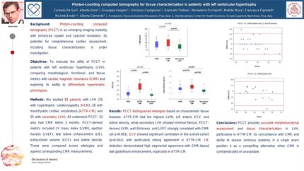
Photon-counting computed tomography for tissue characterization in patients with hypertrophic cardiomyopathy or amyloidosis
European Heart Journal


Abstract
Photon-counting computed tomography (CT) is gaining prominence in cardiac imaging, but its potential for tissue characterization in patients with a hypertrophic phenotype remains to be explored, particularly in comparison with the current gold standard, cardiac magnetic resonance (CMR) imaging.
Consecutive patients with hypertrophic cardiomyopathy (HCM) or amyloid transthyretin cardiomyopathy (ATTR-CM) followed in the outpatient clinic of a referral center for cardiomyopathies were referred to a photon-counting CT as part of a research protocol. A subgroup of these patients underwent also a CMR scan within 3 months from the date of the CT scan, when deemed clinically indicated.
Patients with HCM (n=22) were younger than those with ATTR-CM (n=22; p=0.001), while the percentages of men and women did not differ (p=0.966). On CT scan, patients with HCM had lower values of LV mass (p=0.028) and higher LV ejection fraction (p=0.023), while the extracellular volume (ECV) was lower in patients with HCM (p<0.001). The values of iodine density (a measure of the amount of contrast within the myocardial tissue) did not differ between HCM and ATTR-CM (p=0.101). A subgroup of patients (n=27; n=16 with HCM, n=11 with ATTR-CM) underwent also a CMR scan. Patients with HCM displayed tight correlations between LV mass values from CT and CMR (p<0.001, beta=0.869), and maximal wall thickness (p<0.001, beta=0.969), but no significant correlations between ECV values from the two techniques (p=0.275), or between iodine density and CMR-derived ECV (p=0.274). Patients with ATTR-CM showed significant correlations between LV mass values (p<0.001, beta=0.956) and maximal LV wall thickness (p<0.001, beta=0.990) from CT and CMR, but not between ECV values from the two techniques (p=0.139). Conversely, patients with ATTR-CM displayed a close correlation between iodine density and CMR-derived ECV (p=0.016, beta=0.894).
Photon-counting CT demonstrated strong agreement with CMR for structural parameters (LV mass, maximal wall thickness), supporting its utility for morphologic evaluation. Furthermore, photon-counting CT may hold promise for tissue characterization in patients with ATTR-CM, given the strong concordance between iodine density and CMR-derived ECV.
Contributors

C De Gori
Author

V Castiglione
Author
G Vergaro
Author
F Pignatelli
Author
M Muca
Author
M Occhipinti
Author
M Emdin
Author
A Clemente
Author
You may be interested in