A comprehensive analysis of the normal aortic root using 4D computed tomography
European Heart Journal


Abstract
A precise analysis of the aortic valve and aortic root are essential for valve-sparing surgeries. Preoperative assessment primarily relies on echocardiography. In order to achieve more accurate evaluation, cardiac computed tomography (CT) has emerged as a complementary tool for root analysis. We have previously introduced a novel analysis technique based on 4D (3D+ time) imaging of the normal aortic root.
This study aims to evaluate the dimensions and dynamic changes of the normal aortic root in adult patients using 4D CT.
Using in-house software, 140 cardiac CT of adult patients with a normally functioning tricuspid aortic valve (mean gradient < 15 mmHg, less than 1+ aortic regurgitation on echocardiography) were retrospectively analyzed. A dynamic study of the aortic annulus and other planes of the aortic root was conducted across all 10 temporal phases of the cardiac cycle. Leaflet dimensions were calculated in mid-diastole.
The mean age was 59 ± 14 yrs, with 63% being males. The mean geometric height was 17.5 ± 1.9 mm, the mean effective height was 8.3 ± 1 mm, the mean coaptation height was 4.5 ± 0.7 mm, and the mean commissural height was 23.8 ± 3.7 mm.
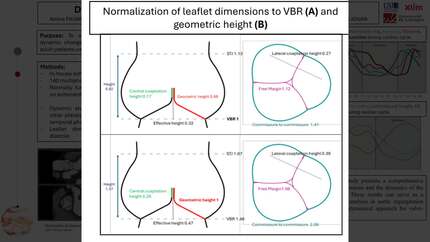
Compared to the left and non-coronary leaflets, the right leaflet exhibited the lowest geometric height (17.1 ± 1.9 mm vs 17.6 ± 2.1 mm and 17.7 ± 1.9 mm, p < 0.001) but the longest free margin (29.1 ± 3.5 mm vs 28.1 ± 3.4 mm and 28.5 ± 3.6 mm, p < 0.001). A positive correlation was observed between the aortic annulus size and the dimensions of the virtual basal ring (VBR), Valsalva sinuses, sinotubular junction (STJ), free margin and geometric height (r= 0.53, 0.81, 0.84, 0.88, 0.67, respectively, p<0.0001). Figure 1 illustrates the normalization of root dimensions in relation to the VBR (A) and to the geometric height (B).
Dynamic analysis revealed a progressive expansion of the aortic root beginning at the level of the VBR (area expansion of 22.1 ± 6.1%), sequentially followed by the aortic annulus (13.2 ± 4.6%), Valsalva sinuses (16.1 ± 7.6%), and STJ (16.4 ± 5.4 %) (Figure 2). During systole, the aortic annulus become more circular (Eccentricity index of 0.45 ± 0.08 in systole vs 0.52 ± 0.09 in diastole, p< 0.001). The planes of VBR and STJ also become more distant (24.33 ± 2 mm in systole vs 23.73 ± 2.17 mm, p=0.04) but more parallel (7.1° ± 2.8 in systole vs 8.9° ± 3.5 in diastole, p<0.001) to one another lining up the outflow tract with the ascending aorta.
This study presents a comprehensive analysis of the dimensions and the dynamics of the normal aortic root. These results can serve as a reference for valve analysis in aortic regurgitation and hence guide a dynamical approach for valve-sparing surgeries. Normalization of root dimensions


You may be interested in