Imaging findings and clinical analysis of cardiac hemangioma
European Heart Journal


Abstract
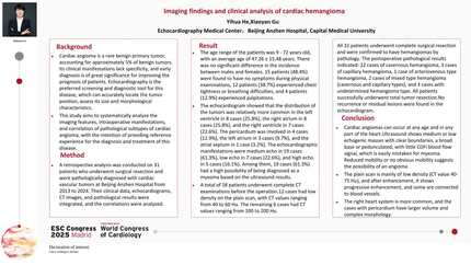
Cardiac hemangioma, a rare benign tumor representing ~5% of benign cardiac tumors, often lacks specific symptoms. Echocardiography is key for screening/diagnosis, enabling precise tumor localization and morphological evaluation. This study analyzes imaging features, surgical observations, and pathological correlations to enhance diagnostic and therapeutic strategies.
A retrospective analysis was conducted on 31 patients who underwent surgical resection with pathological confirmation of cardiac hemangioma at our Hospital from 2013 to 2024. Clinical data, echocardiographic findings, CT imaging, and pathological results were integrated to analyze correlations.
Patients aged 9–72 years (mean 47.3±15.5), no gender predominance. Asymptomatic (48.4%, 15/31) detected incidentally; symptomatic presentations: chest tightness/dyspnea (38.7%, 12/31), palpitations (12.9%, 4/31).
Tumor distribution included the left ventricle (8 cases, 25.8%), right atrium (8 cases, 25.8%), right ventricle (7 cases, 22.6%),less common in pericardium, left atrium and atrial septum.Echocardiographic features included medium echogenicity (19 cases, 61.3%), hypoechoic masses (7 cases, 22.6%), and hyperechoic masses (5 cases, 16.1%). Notably, 19 cases (61.3%) were initially suspected as myxomas.
Right ventricle (7 cases): Six pedunculated masses with reduced mobility (suspected myxomas), one thrombus-like mass without mobility. Right atrium (8 cases): Five located in the interatrial septum, 3 in the lateral wall; 6 suspected as myxomas (only 1 with significant mobility).Left ventricle (8 cases): Four in the interventricular septum, 2 in the inferoposterior wall, 2 on the anterior mitral leaflet; 6 suspected as myxomas with mild mobility, 2 thrombus-like masses. Left atrium (3 cases): Located in the fossa ovalis (1 mobile mass suspected as myxoma) and left atrial appendage.
Non-contrast scans revealed soft-tissue masses with progressive enhancement post-contrast. Twelve cases showed hypodense lesions (CT value: 40–60 HU), while 6 exhibited hyperdense lesions (CT value: 100–200 HU).
All 31 patients underwent complete tumor resection. Pathological analysis confirmed 22 cavernous hemangiomas, 3 capillary hemangiomas, 1 arteriovenous hemangioma, 2 mixed-type (cavernous and capillary) hemangiomas, and 3 unclassified subtypes. Postoperative echocardiographic follow-up (mean: 24 months, range: 6–60 months) showed no recurrence or residual lesions.
Cardiac hemangiomas can arise in any cardiac region at any age, often asymptomatically, but show right heart predominance. Echocardiography reveals well-defined medium/hypoechoic masses, often broad-based or pedunculated and mistaken for myxomas; limited mobility hints at hemangioma. While multimodal imaging aids preoperative accuracy, histopathology confirms diagnosis. Early surgery is advised for suspected cases, with routine echo follow-up to detect recurrence.


You may be interested in