Echocardiographic AMYLI score in systemic light-chain amyloidosis: clinical relevance and risk stratification
European Heart Journal


Abstract
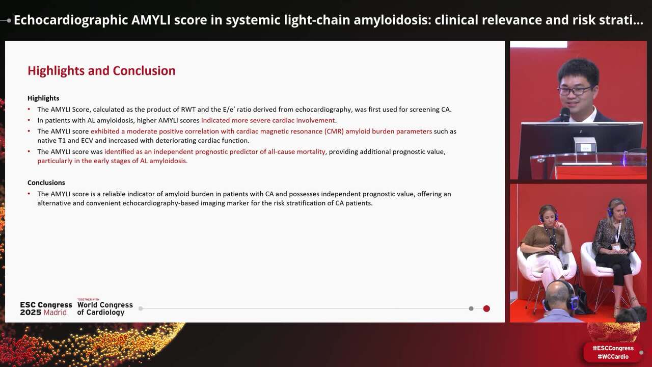
While the AMYLoidosis Index (AMYLI) has been extensively utilized for screening cardiac amyloidosis (CA). However, the relationship between this parameter and amyloid burden, as well as its prognostic value, remains elusive.
This study aimed to investigate the association between the AMYLI Score and amyloid burden and its prognostic value in patients with CA.
This single-center, prospective, observational study was conducted at our hospital from November 2011 to September 2023. A total of 321 patients with biopsy-proven AL amyloidosis who underwent both echocardiography and cardiac magnetic resonance (CMR) imaging were enrolled. The AMYLI score was calculated via transthoracic echocardiography as the product of relative wall thickness (RWT) and E/e′. Lastly, parameters reflecting amyloid burden, such as late gadolinium enhancement, native T1, and extracellular volume (ECV), were calculated via CMR.
The AMYLI score exhibited a moderately positive correlation with CMR parameters associated with amyloid burden, such as native T1 value (r = 0.42, P < 0.001) and ECV (r = 0.57, P < 0.001). During a median follow-up of 42 (interquartile range: 35-49) months, 205 patients died. Meanwhile, an AMYLI score ≥ 10.08 demonstrated a strong predictive value for all-cause mortality [hazard ratio (HR) 2.40, 95% confidence interval (CI): 1.74-3.32, P < 0.001]. After adjusting for clinical (HR 1.56, 95% CI: 1.11-2.21, P = 0.009), biochemical (HR 1.55, 95% CI: 1.10-2.18, P = 0.009), echocardiographic (HR 1.83, 95% CI: 1.31-2.57, P = 0.037), CMR imaging-related (HR 1.51, 95% CI: 1.02-2.25, P = 0.037), and therapeutic (HR 1.50, 95% CI: 1.07-2.11, P < 0.001) factors, an AMYLI score ≥ 10.08 remained an independent prognostic factor.
The AMYLI score is a reliable indicator of amyloid burden in patients with CA and possesses independent prognostic value, offering an alternative and convenient echocardiography-based imaging marker for the risk stratification of CA patients. graphic abstract examples


You may be interested in