Detection of high-risk severe aortic stenosis using coronary flow pattern
European Heart Journal


Abstract
Symptomatic severe aortic stenosis (AS) has a poor prognosis and requires therapeutic intervention. However, identifying symptoms in elderly patients can be challenging. Therefore, objective indicators are needed to detect high-risk AS. Previous studies have shown that left ventricular global longitudinal strain (GLS) and B-type natriuretic peptide (BNP) serve as indicators of high-risk AS (1–2).
In patients with AS, increased systolic wall stress due to elevated afterload reduces systolic coronary flow, often leading to systolic coronary flow reversal (SFR) in the epicardial coronary arteries (Figure 1).
This study aimed to evaluate whether SFR can serve as an indicator of high-risk AS. Using transthoracic echocardiography (TTE), we measured coronary flow in patients with severe AS and assessed the relationship between the presence or absence of SFR and GLS and BNP levels.
Between July 2023 and February 2025, patients undergoing TTE for AS evaluation were prospectively enrolled. The severity of AS and left ventricular function were assessed based on ESC guidelines. Additionally, TTE was used to measure distal left anterior descending coronary artery flow. SFR was defined as the presence of a reversed coronary flow component in early systole.
Patients with severe AS were classified into SFR+ and SFR− groups based on the presence or absence of SFR in coronary flow measurements. The inclusion criterion was severe AS, while the exclusion criteria were the inability to assess coronary flow and non-severe AS.
Of the 89 enrolled cases, 9 were excluded due to poor coronary imaging, and 24 were excluded due to non-severe AS. The remaining 56 patients with evaluable coronary arteries and severe AS were classified into the SFR+ group (24 cases) and the SFR− group (32 cases).
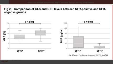
A comparison of echocardiographic data between the two groups revealed no significant differences in left ventricular indicators such as left ventricular mass index, left ventricular ejection fraction, and E/e’. However, GLS was significantly lower in the SFR+ group compared to the SFR− group (11.9 ± 3.4% vs. 15.5 ± 3.3%, P<0.01).
Regarding AS severity, the aortic valve peak velocity was significantly higher in the SFR+ group than in the SFR− group (481 ± 70 cm/s vs. 433 ± 33 cm/s, P<0.01). In blood test results, BNP levels were significantly higher in the SFR+ group than in the SFR− group (526 [89–1184] pg/ml vs. 156 [82–266] pg/ml, P=0.01) (Figure 2).
The presence of SFR may serve as a simple indicator for detecting high-risk severe AS. Incorporating coronary flow measurement into routine clinical echocardiography may facilitate simple and effective risk stratification of severe AS.
Contributors

M Fujimoto
Author
K Mukai
Author
H Ohashi
Author
A Kunimura
Author
Y Nakano
Author
H Ando
Author
T Amano
Author
You may be interested in