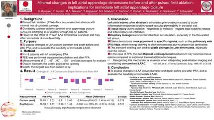
Minimal changes in left atrial appendage dimensions before and after pulsed field ablation: implications for immediate left atrial appendage closure
European Heart Journal


Abstract
Pulsed field ablation (PFA) has emerged as a promising modality for pulmonary vein isolation due to its high selectivity for myocardial tissue and reduced risk of collateral damage. Unlike conventional thermal ablation techniques, PFA minimizes the risk of esophageal injury, phrenic nerve damage, and pulmonary vein stenosis. Recently, the combination of catheter ablation and left atrial appendage (LAA) occlusion has been shown to be beneficial, particularly in patients with atrial fibrillation at high thromboembolic and bleeding risk. However, the effects of PFA on LAA dimensions remain largely unexplored.
Given that myocardial edema can occur post-ablation, potentially altering atrial and appendage structure, a better understanding of these changes is crucial to ensuring the safety and efficacy of simultaneous procedures. If PFA significantly alters LAA geometry, it could affect the feasibility of immediate LAA closure or influence device selection.
This study aimed to evaluate changes in LAA ostium and depth dimensions before and after PFA to assess its impact on the immediate feasibility of LAA closure.
We prospectively analyzed eight patients who underwent PFA for atrial fibrillation. Transesophageal echocardiography (TEE) was performed intraoperatively to measure LAA ostium diameter and depth before and immediately after PFA. Measurements were taken at four standard imaging angles (0°, 45°, 90°, and 135°), and the average values across these angles were used for analysis.
LAA ostium diameter was defined as the maximal width at the ostium, while LAA depth was measured from the ostium to the tip of the appendage along its longest axis. Statistical comparisons were performed using paired t-tests, and significance was defined as p < 0.05.
Pre-PFA ostium diameters ranged from 16.1 mm to 21.5 mm (mean 18.89 ± 2.03 mm), while post-PFA diameters ranged from 15.7 mm to 20.9 mm (mean 18.21 ± 2.00 mm). The mean reduction in ostium diameter was -0.68 mm (95% CI: [-1.46, +0.10], p = 0.078).
LAA depth ranged from 14.6 mm to 20.7 mm pre-PFA (mean 16.96 ± 2.03 mm) and 13.6 mm to 17.6 mm post-PFA (mean 15.96 ± 1.48 mm). The mean reduction in depth was -0.997 mm (95% CI: [-2.33, +0.34], p = 0.121).
Neither LAA ostium diameter nor depth exhibited a statistically significant change post-PFA. The distribution of changes was consistent across different imaging angles, suggesting minimal regional variation in structural response.
These findings suggest that PFA does not significantly alter LAA geometry, supporting the safety of immediate LAA closure post-ablation. The minor, nonsignificant reductions in ostium and depth dimensions may reflect transient edema without substantial remodeling. Given the small sample size, further studies with larger cohorts are warranted to confirm these observations and assess potential implications for LAA closure device sizing and placement. LAA ostium diameter before and after PFA LAA depth before and after PFA
Contributors


You may be interested in