Detection of vegetations in transesophageal echocardiographic images of patients with infective endocarditis using artificial intelligence
European Heart Journal


Abstract
Infective endocarditis (IE) is a serious, life-threatening condition associated with severe complications, often leading to the formation of vegetations, on the heart valves [1]. These vegetations are correlated with cerebral embolisms [2], and the probability of embolization is influenced by vegetation features such as size and shape [3], which can be difficult to measure [4]. Current models to estimate that risk have shown little predictive power [3]. This study introduces an artificial intelligence (AI) system designed to detect IE-associated vegetations within the left heart chambers in Transesophageal Echocardiography (TEE) images.
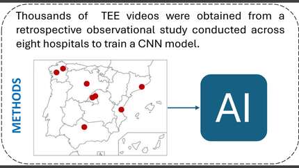
Retrospective observational study across 8 hospitals with two distinct phases. In the first phase, data from 203 patients were manually annotated and used to train an AI model to receive a TEE still image as an input, and, if any vegetation is found, output the coordinates of detected vegetations together with a confidence level. Model’s performance was evaluated using traditional frame-level accuracy metrics, assessing its ability to detect vegetations in individual echocardiographic images.
In the second phase, a clinical validation test was performed to assess the model’s ability to distinguish between patients with and without vegetations in complete TEE video sequences. This test involved a separate cohort of 60 patients (30 with vegetations and 30 without). Instead of evaluating individual frames, the frame-based AI model developed in the first phase was sequentially applied, frame-by-frame, to each TEE video, and the output of each frame aggregated to output a prediction on the presence or absence of vegetations across the video.
In the first phase, the model achieved promising performance metrics with precision=0.81 and recall=0.72 for vegetation detection in individual still images. In the second phase, clinical validation demonstrated the model’s robust diagnostic capability to identify patients with vegetations when presented with entire video sequences, as evidenced by an area under the receiver operating characteristic curve (AUC) of 0.92.
The algorithm achieved high-performance metrics detecting vegetations and identifying patients with vegetations, which can be useful to automate vegetation measurements and facilitate and accelerate IE diagnosis by non-expert cardiologists. Detection vs. ground truth Methodology scheme
Contributors

L Llamas-Fernandez
Author
M Carrasco-Moraleja
Author
I Gomez
Author
C Gonzalez-Juanatey
Author
C Pidone
Author
M Anguita-Sanchez
Author
J C Lopez-Azor
Author
M A Arnau Vives
Author
A Vallejo-Sevillano
Author
J A San Roman
Author
C Baladron
Author
You may be interested in