Cell painting based high-content phenotypic screening to elucidate kinase signaling pathways in ibrutinib-mediated atrial fibrillation
European Heart Journal Supplements


Abstract
One goal of Precision Cardio-Oncology is to identify and ultimately clinically exploit a fundamental understanding of shared signaling pathways in cardiovascular disease and oncologic processes. One example is ibrutinib, which inhibits BTK (Bruton’s tyrosine kinase) and CSK. While ibrutinib has revolutionized treatment for B-cell malignancies, it has increased the incidence of atrial fibrillation (AF) compared with conventional chemotherapy.
Here we report the use of high-content imaging and phenotypic screening of human iPSC-derived cardiomyocyte models to inform mechanisms underlying this proarrhythmic process.
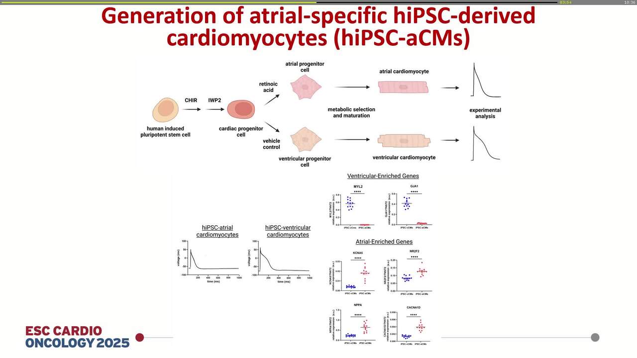
Studies were performed in human atrial-specific cardiomyocytes (hiPSC-aCMs) derived from population control induced pluripotent stem cells. Monolayers of hiPSC-aCMs were plated onto 96-well plates and treated with increasing concentrations of the BTK and CSK inhibitor ibrutinib or DMSO-vehicle control. We have previously shown that ibrutinib treatment of hiPSC-aCMs results in proarrhythmic behavior. After 72 hours of ibrutinib treatment, in situ staining with the Cell Painting assay was performed. This allows morphologic profiling by simultaneously multiplexing multiple different fluorescent dyes to label different cellular components (e.g. mitochondria, nuclei, endoplasmic reticulum, and cytoskeleton), including mitochondrial structure. Measurements of mitochondrial intensity, texture, shape, size, and relation to neighboring cellular structures were recorded and analyzed in relation to treatment with ibrutinib or vehicle control.
Treatment of hiPSC-aCMs with ibrutinib resulted in dose-dependent changes in 34 parameters of mitochondrial structure, including changes in mitochondrial intensity, texture, shape, size, and relation to neighboring cellular structures. For example, a statistically significant dose-dependent response following ibrutinib treatment was observed in mean mitochondrial intensity (p<0.001 and p<0.0001 for ibrutinib 1 µM and 10 µM, respectively, Figure 1). 3D-principle component analysis (PCA) of all 34 measured mitochondrial parameters showed significant differences (p<0.0001) in mitochondrial structure between ibrutinib and vehicle-treated hiPSC-aCMs.
Ibrutinib exposure causes marked changes in multiple indices of mitochondrial structure, implicating altered mitochondrial function in the development of ibrutinib-related AF. Decreased mitochondrial oxidative phosphorylation and gene expression have been previously shown in hiPSC-aCMs exposed to ibrutinib, providing further biological relevance to these observed phenotypic changes.
Contributors

You may be interested in