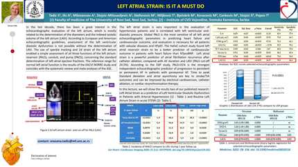
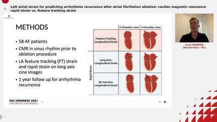
Left atrial strain as a predictor of atrial fibrillation in hypertrophic cardiomyopathy
EP Europace Journal


Abstract
Atrial fibrillation (AF) stands as the prevailing arrhythmia across all forms of cardiomyopathies. The occurrence of AF in individuals with hypertrophic cardiomyopathy (HCM) is 4 to 6 times more frequent compared to the general population of the same age. Patients (P) with HCM and FA are at higher risk of adverse outcomes. To date, there is not a widely validated model that can predict the risk for development of AF in P with HCM. We aimed to study the role of left atrial (LA) myocardial deformation imaging as a toll for predicting FA development, in a population with HCM.
P with HCM accompanied at our Cardiomyopathies Center who had no palpitations or documented AF at the time of echocardiographic evaluation were included. During follow-up, AF was established as an atrial tachyarrhythmia with uncoordinated atrial electrical activation lasting more than 30 seconds. For evaluation of outcomes, our cohort was divided into two groups (with and without AF). For the group with AF, the last transthoracic echocardiogram (TTE) performed before the diagnosis of AF was considered, for the group without AF, a TTE was analyzed in P with a similar follow-up duration. LA deformation imaging using two-dimensional speckle tracking echocardiography was performed according to the consensus document of the EACVI/ASE/Industry Task Force to standardize deformation imaging.
Forty-nine P with HCM (age 70.60±12.0, 43% male sex) were included. Twenty-six (53%; age 63.3±13.11, 30.4% male sex) developed AF during the follow-up (FU) (mean FU of 29,5± 25 months). The groups had similar baseline clinical and demographic characteristics, and no significant differences were registered when comparing clinical aspects or regular medication. When analyzing the LA strain, there were no significative differences between the reservoir or conduit phases of the LA cycle, in either apical four chamber (A4C) view, apical two chamber (A2C) view or biplane analysis. The contraction phase of the LA cycle was significantly different between the two groups (p=0.02 for A4C, p=0.04 for A2C and p=0.05 for biplane). The ROC curves were drawn, with an area under the curve of 0.665 for A4C, 0.698 for A2C and 0.673 for biplane. Based on ROC curve analysis, an optimal cut-off point of -8.5% for sensitivity and specificity in the contraction phase of the LA cycle strain was determined. After Cox regression analysis, P with a value of LA strain for the contraction phase ≤-8.5% had a higher risk of developing AF (hazard ratio [HR] 4.29; 95% confidence interval [CI] 1.25-14.74, p value 0.021).
In our cohort of HCM P, atrial strain, particularly the contraction phase of the LA cycle, appears to be a valuable predictor for the development of AF. LA strain in patients with HCM Hazard Curve for AF development
Contributors

I Ferreira Neves
Author
M Caetano Coelho
Author
M Marques Antunes
Author
P Garcia Bras
Author
A Ferreira
Author
I Cardoso
Author
J M Viegas
Author
I Almeida
Author
A Fiarresga
Author
P Silva Cunha
Author
R Cruz Ferreira
Author
M Martins Oliveira
Author
S Aguiar Rosa
Author
You may be interested in