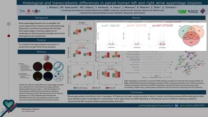
Histological and transcriptomic differences in paired human left and right atrial appendage biopsies
EP Europace Journal


Abstract
Atrial appendage biopsies form a valuable, but scarce opportunity to study atrial pathology in human cardiovascular disease. Despite their frequent use, systematic comparisons between left and right atrial appendages is lacking, largely due to challenges of obtaining paired samples and clinical heterogeneity among patients.
To compare histology and gene expression in paired left and right atrial tissue samples.
Paired left (LA) and right (RA) atrial tissue samples were obtained from cardiovascular surgery patients (bypass graft/valvular surgery, n=92) in the RACE V tissue bank study. Histological and transcriptomic differences between LA/RA were studied in patients without AF history (n=52), with paroxysmal AF (n=16) and persistent AF (n=25). Biopsies were stained (WGA/CD31/Vimentin) to quantify endomysial and overall fibrosis, fibroblast density, myocyte diameter, capillary size and density. Histological differences were tested using paired t.tests. Transcriptomic patterns were evaluated using bulk RNA-sequencing.
RA samples consistently contained more overall and endomysial fibrosis than LA samples independent of rhythm group (Fig.1). In patients with a history of AF, LA samples had larger cardiomyocytes. Gene expression analyses revealed 2002 differentially expressed transcripts in patients without AF history, and 5770 in persistent AF patients. Independent of rhythm history, the strongest LA genes were PITX2 and the adjacent PANCR, while BMP10 was the strongest RA gene. Enriched processes differentiating both atria were related to heart morphogenesis, regulation of blood pressure and BMP signaling, independent of rhythm history. Response to fibroblast growth factor, connective tissue development and heart contraction were specifically enriched in AF patients.
Right atria are more fibrotic and differ in gene expression patterns from left atria, potentially contribution to differences in susceptibility to atrial pathology.
Contributors

J Winters
Author
M J Kawczynski
Author
M D Gilbers
Author
S Verheule
Author
A Isaacs
Author
J Maessen
Author
B Maesen
Author
E Bidar
Author
U Schotten
Author
You may be interested in