Repolarization and depolarization abnormalities are preferentially impaired by ajmaline in Brugada patients compared to controls
EP Europace Journal


Abstract
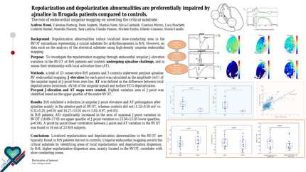
Repolarization hypothesis states that a transmural repolarization dispersion in right ventricular outflow tract (RVOT) determines type-1 ECG phenotype in Brugada syndrome (BrS). So far only few evidences support it, whilst depolarization impairment has been widely described.
To investigate the changes in depolarization mapping through endocardial unipolar J-elevation mapping of RVOT in BrS patients and in controls undergoing ajmaline and their relations with activation time (AT), marker of local depolarization.
Consecutive BrS patients with spontaneous type-1 ECG pattern and controls underwent RV endocardial mapping with the CARTO3 system. Mapping in both groups was performed before (pre) and after (post) ajmaline administration as none expressed a baseline type-1 ECG pattern. Data were exported and converted into Matlab format using OpenEP. J-elevation for each point was calculated as the amplitude in mV of the unipolar signal at J-point from zero-line. For each point AT was defined as the difference between local depolarization (minimum -dV/dt of the unipolar signal) and surface ECG depolarization (minimum of V2). Corresponding pre and post points were selected; patients having <200 corresponding points were discarded. In each patient, pre and post AT and J-elevation maps were created; a region of interest (ROI) covering the RVOT was selected based on the tricuspid valve position. For each ROI point delta-J and delta-AT were calculated as the difference between post and pre, and interpolated to obtain RVOT differential maps. A point-by-point correlation analysis was performed between delta-J and delta-AT. A maximum and a minimum variation area (ROImax and ROImin, respectively) within RVOT were identified for both delta-J and delta-AT.
24 BrS patients and 2 controls were included (1092±633 average corresponding points); all BrS patients presented type-1 ECG after ajmaline, while no control did. Examples of RV maps, ROI selection and RVOT pre, post and differential maps can be seen in Figure 1. In BrS, ROImax delta-J was significantly higher with respect to ROImin (1.68 vs 0.6 mV, p<0.001) while delta-AT ROImax was significantly longer compared to delta-AT ROImin (24.1 vs 8.72 ms, p=0.004). In controls no differences between ROImax and ROImin were observed for delta-J, nor for delta-AT. (Figure 2). In BrS population, a point-by-point correlation between delta-J and delta-AT was present in patient BrS4 (Pearson R coefficient=0.61, p<0.001) and in patient BrS20 (R=0.53, p> 0.001).
Localized repolarization and depolarization abnormalities in the RVOT are typically found in BrS patients but not in controls. High-density unipolar endocardial mapping pre and post-ajmaline unveils the critical electrophysiological substrate unmasking local repolarization and depolarization dispersion area. In BrS, higher repolarization dispersion area, located in the RVOT, correlates to slow-conducting zones. Methods Results
Contributors

A Rossi
Author
V Hartwig
Author
S Latrofa
Author
P Seghetti
Author
S Garibaldi
Author
M Nesti
Author
L Panchetti
Author
G Mirizzi
Author
U Startari
Author
N Zaurino
Author
M S Morelli
Author
C Passino
Author
M Emdin
Author
M Piacenti
Author
A Giannoni
Author
You may be interested in