The use of LGE-derived 3D scar metrics to predict ICD therapy in ischemic cardiomyopathy patients
EP Europace Journal


Abstract
Implantable cardioverter-defibrillators (ICDs) play a vital role in the management of ventricular arrhythmias (VAs). To improve arrhythmia risk-stratification, late gadolinium enhancement (LGE) imaging can be used to identify and characterize the underlying arrhythmogenic substrate.
This study aims to evaluate the relationship between LGE-derived quantitative scar metrics and appropriate ICD therapy.
Patients with ischemic cardiomyopathy who underwent LGE imaging prior to ICD implantation between 2017 and 2019 were retrospectively identified. Scar characteristics were quantified using ADAS LV (ADAS LV Medical, Barcelona, Spain). Additional interface metrics were obtained using customized scripts. Magnitude (MAG) images were analysed and, when available, compared with phase-sensitive inversion recovery (PSIR) images to assess differences in scar quantification. Follow-up data was analysed to identify patients that received appropriate ICD therapy.
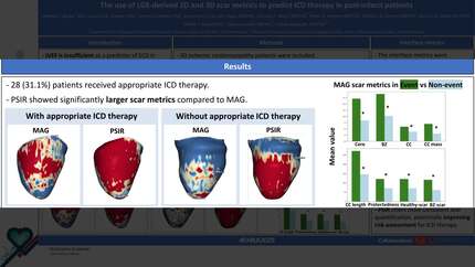
A total of 90 patients were included. Over a median follow-up period of 63 months (IQR: 48-74 months), 28 (31%) received appropriate ICD therapy.
MAG analysis revealed significant differences in core scar (8.37±5.96g vs. 17.1±7.61g, p<0.001), border zone (BZ) (10.18±5.78g vs. 19.03±6.77g, p<0.001), conduction corridor (CC) mass (3.03±3.11g vs. 6.95±3.66g, p<0.001), CC number (3.85±3.28 vs. 5.82±3.16, p=0.005), CC length (146.19±150.05mm vs. 271.36±138.85mm, p<0.001) and CC protectedness (71.5±89.83mm vs. 120.66±89.14mm, p<0.001) between patients with and without appropriate therapy. Furthermore, the interface between healthy myocardium and scar (70.64±39.77cm2 vs. 121.00±40.23cm2, p<0.001), as well as the interface between BZ and scar (61.61±35.07cm2 vs. 116.12±37.62cm2, p<0.001) was significantly larger in patients with appropriate ICD therapy.
Additionally, with 80 cases available for comparison, significant differences were observed between MAG and PSIR analyses. PSIR showed larger core (11.15±7.75g vs. 18.83±11.37g, p<0.001), BZ (12.57±6.85g vs 23.12±12.85g, p<0.001), CC mass (4.16 ± 3.80g vs. PSIR: 5.58 ± 4.91g, p=0.038) and CC length (186.08 ± 160.31mm vs. 226.85 ± 169.23mm, p=0.041), although the total CC number (p=0.26) and CC protectedness did not differ significantly between the two reconstruction methods (p=0.079). Moreover, the interface between healthy myocardium and scar (84.30±45.56 cm2 vs. 112.47±60.50cm2, p<0.001) and the interface between BZ and scar (89.36±95.87cm2 vs. 100.17±87.19cm2, p<0.001) were significantly different for MAG and PSIR.
LGE-derived quantitative scar metrics are predictors of appropriate ICD therapy in patients with ischemic cardiomyopathy. The differences observed between PSIR and MAG suggest that PSIR may offer more detailed and accurate scar quantification, potentially improving risk assessment for ICD therapy.
Contributors

J C Burger
Author
L H G A Hopman
Author
F O Campos
Author
A C Van Der Lingen
Author
C P Allaart
Author
P G Postema
Author
M J B Kemme
Author
M J W Gotte
Author
M J Bishop
Author
V P Van Halm
Author
P Bhagirath
Author
You may be interested in