Unmasking conduction block zones associated with scar-related ventricular tachycardia circuits by the peak frequency mapping and three-dimensional cardiac magnetic resonance imaging
EP Europace Journal


Abstract
Catheter ablation (CA) for ventricular tachycardia (VT) often fails to identify the arrhythmogenic substrate and circuit. The peak frequency (PF) mapping is a useful indicator for distinguishing far-field and near-field electrograms mixed in the critical isthmus of conduction [1, 2]. It has been reported that advanced cardiac imaging prior to the CA procedure can enhance treatment efficacy and contribute to improved successful ablation rate [3, 4]. However, the relationship between PF and cardiac magnetic resonance (MR) imaging findings in identifying the critical isthmus of conduction remains insufficiently elucidated.
This study aimed to investigate the association between cardiac MR imaging findings and the peak frequency mapping and to determine its efficacy of VT ablation.
From 2023 to 2024, 7 consecutive cases with sustained monomorphic VT (6 men, age 55±9 years) who underwent CA were enrolled, with PF maps retrospectively generated. Cardiac MR was performed in 6 out of 7 cases. The imaging data were analyzed using 3-dimensional imaging software to detect the target arrhythmogenic substrate of VT and were then merged with the mapping system.
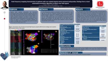
In the 7 cases, 5 had ischemic cardiomyopathy. VT mapping was successfully performed in 5 out of the 7 cases. In the remaining 2 cases, the clinical VTs were unmappable due to hemodynamic instability or non-inducibility. Substrate mapping during sinus rhythm, right ventricular, and left ventricular pacing was evaluated using a combination of isochronal late activation mapping and activation mapping based on the near-field annotation algorithm in all cases. U-shaped conduction blocks during substrate mapping were identified during one or more pacing conditions in 5 out of 7 cases, and the conduction blocks coincided with the VT isthmus in 4 of these cases. In the remaining case of ischemic cardiomyopathy, clinical VTs were unmappable. However, the interior of the U-shaped conduction block corresponded to the good pace mapping score sites, which matched the perfusion-metabolism mismatch areas identified by radioisotope imaging, suggesting contribution of the myocardial ischemia as a VT arrhythmogenic substrate (Figure 1). The PF in these specific regions related to VT critical sites was high (539.2±98.3Hz), with the highest PF being 716Hz and the lowest PF being 426Hz during substrate mapping (Figure 2). VT could be terminated with minimal ablation in these regions in 3 out of 7 cases. These regions that participated in VT circuit coincided with the borders of the dense scar in the total scar in 5 out of 6 cases (83%) evaluated by cardiac MR, suggesting that the boundaries forming the VT circuit were related to scar heterogeneity.
The optimal target ablation sites for scar-related VT can be revealed by using a combined approach of the PF analysis during substrate mapping with cardiac MR based scar distribution and heterogeneity 3-dimensional imaging.
Contributors

M Hachisuka
Author
Y Iwasaki
Author
S Okajima
Author
N Ito
Author
S Kobayashi
Author
R Mimuro
Author
Y Fujimoto
Author
H Hayashi
Author
H Murata
Author
Y Aizawa
Author
K Yodogawa
Author
W Shimizu
Author
K Asai
Author
You may be interested in