Impact of myocardial scar pattern on transmural repolarisation gradients in non-ischaemic VT
EP Europace Journal


Abstract
Repolarisation heterogeneities play an important role in ventricular arrhythmogenesis, yet detailed high-resolution assessment is challenging. In vivo research of repolarisation dynamics in VT substrate is predominantly limited to ischemic substrate and assessed on a single surface. Less is known about their role in non-ischemic cardiomyopathies (NICM) with frequently more complex mid- and subepicardial scar pattern.
Evaluate and characterise transmural repolarisation gradients (TMG) in a clinical cohort of patients with complex non-ischemic scar patterns undergoing combined endo-epicardial electro-anatomical substrate mapping.
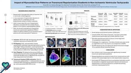
Methodology: Patients with NICM and prior high-density endo-epicardial substrate mapping as part of a clinically indicated VT ablation procedure and preprocedural cardiac MRI to establish scar pattern were retrospectively reviewed. Unipolar repolarisation substrate maps were generated of both endo- and epicardium using the TurboMap feature. Local repolarisation Time (RT) was defined from beginning of surface QRS to the maximum +dVdt of the T wave on unipolar EGMs according to the Wyatt method. 3D delayed enhancement CMR scar models were reconstructed and co-registered with the EAM-RT map using anatomical landmarks. Sites of transmural, midmyocardial and subepicardial scar were marked. Transmural gradients were assessed by subtraction of corresponding epi- and endocardial EGM-RTs (in ms) of manually selected EGM pairs opposing each other in areas of scar, adjacent to scar and remote healthy myocardium.
15 patients (80% male, 47±15years, LVEF 38±14%, aetiology: 60% ACM, 20% Idiopathic DCM, 20% post-myocarditis) were studied. All had left-ventricular non-ischemic scar. Local RTs were longest over areas of transmural scar followed by non-transmural scar and shorter in healthy myocardial segments (ANOVA p <0.001, post-hoc pairwise <0.001 respectively).
Transmural gradients across segments were significantly different (p<0.001), including post-hoc pairwise comparison for transmural scar vs healthy (p=0.010) and non-transmural scar vs healthy segments (p <0.001) (Table 1). Transmural gradients were largest at sites of non-transmural scar (subepicardial+/-midmyocardial) and greater than 2-fold higher (+111%) than TMGs in healthy myocardium. Transmural scar and border zone had +59% higher gradients than healthy tissue but less than in non-transmural scar. In the remote healthy segment, a physiological epi-to endocardial RT gradient was observed which reversed to endo-to-epicardial gradient at sites of scar and border zone - reversal was most prominent at sites of non-transmural (epi-mid) scar.
In patients with non-ischemic scar and ventricular arrhythmias, significantly increased transmural repolarisation gradients are present with steepest gradients at sites of non-transmural scar with reversal of the physiological epi-to-endo repolarisation sequence. Endo-epicardial RT Maps and EGM-Pairs Endo-Epicardial RTs and TMGs in NICM
Contributors

J B Tonko
Author
E Cabrera-Borrego
Author
P Sanchez-Millan
Author
J Jimenez-Jaimez
Author
A Chow
Author
P Lambiase
Author
You may be interested in