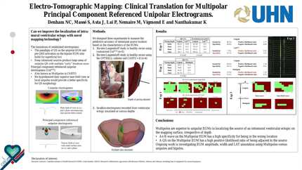
Electrotomographic mapping: clinical translation for multipolar principal component referenced unipolar electrograms
EP Europace Journal


Abstract
Focal ventricular arrhythmias from an intramural location pose a significant challenge to map and ablate. The paradigm of QS on the unipolar electrogram (EGM) and pre-QRS activation on the bipolar EGM only holds for superficial foci and is lost for deeper sources owing to mass depolarization of myocardium leading to a dual breakout pattern over a large surface area. We recently reported multipoles with principal component referenced unipolar electrograms (UniPCR) have a superior near-field view and demonstrated a lower fractional area of QS EGMs with a deep pacing source.
We hypothesized multipoles with UniPCR would be a better predictor of intramural source location irrespective of depth on the surface of the heart by acting as a local unipolar EGM with a better specificity for QS morphology.
Five explanted healthy swine hearts were used in a Langendorff perfused heart model. Mapping data was collected using a custom-made, 2mm spaced, 56-electrode rectangular array placed on the epicardium of the left ventricle. Pacing was performed from the center of the array, varying the intramural stimulation source depth by plunge needle in 1mm increments from 6mm (deepest) to 3mm (shallowest). A comparison of unipolar versus UniPCR EGMs was performed to measure the predictive accuracy of the source location including amplitude, width, QS% and fractional area of QS. Data are presented as mean±SEM.
The percentage of EGMs displaying a QS morphology was lower in UniPCR vs. Unipolar EGMs at all depths (p<0.001; Table). This produced a smaller fractional area of QS on the array irrespective of source depth (p<0.001). A representative example from one swine is shown in the figure. UniPCR EGMs had a smaller amplitude and narrower width than unipoles (both p<0.001), however there was no difference in the amplitude or width of UniPCR EGMs across the different depths of the pacing source (p=0.957 and p=0.372 respectively).
Electrodes on the array were subdivided into 1st neighborhood (four electrode square around source), 2nd neighborhood (twelve electrode square around 1st neighborhood) and remote (all remaining electrodes). The percentage of EGMs displaying a QS morphology was lower in UniPCR vs. Unipolar EGMs at all depths in the 1st neighborhood (p<0.001), 2nd neighborhood (p<0.001) and remote (p<0.001). A non-QS signal on UniPCR had much better specificity for being at an electrode outside the 1st neighborhood (unipolar: 60.4% vs. UniPCR: 91.5%).
Multipoles with UniPCR improves the ability to map an intramural focus by creating a narrower area of QS on the mapping surface due to reduced far-field influence on the EGM. Traditional unipoles show a larger area of QS due to far-field summation. Multipoles allows for effective source localization on the surface irrespective of depth and has the potential to improve targeted ablation of focal ventricular arrhythmias. QS vs. R wave maps Characteristics of EGMs

You may be interested in