Characterization of static and dynamic depolarization and repolarization characteristics in the short-QT syndrome
EP Europace Journal


Abstract
The short-QT syndrome (SQTS) is a rare cardiac disorder associated with abnormally short repolarization duration and increased risk of ventricular tachyarrhythmia (VT). In-vivo depolarization and repolarization patterns in patients with SQTS are unknown.
First characterization of epicardial depolarization and repolarization in SQTS using noninvasive ECG imaging (ECGi).
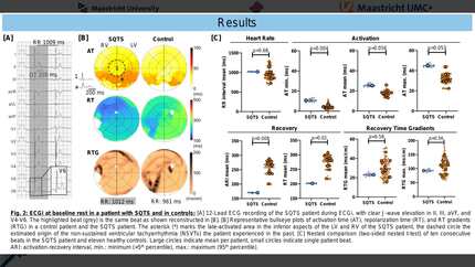
ECGi was performed in a 54-year-old male SQTS patient (QTc 318 ms, inferolateral J-wave elevation, SQTS score 4, no causal mutation identified) with a history of syncope and long-coupled apical non-sustained VT, treated with metoprolol (Figure 1). Ten consecutive sinus beats were recorded at rest, directly after air cycling, and five minutes after exercise. Results were compared to a control group of n=11 healthy controls (27% male, 59±6 years, QTc 415±18 ms). ECGi consisted of a 224-electrode body-surface potential map and a contrast-enhanced CT scan to determine torso and heart geometry. Local activation (AT) and recovery times (RT) were derived from reconstructed epicardial unipolar electrograms. Data are shown as mean±SD.
RR intervals of the SQTS patient and controls were comparable (1019±9 vs. 955±157 ms, p=0.68). The initial phase of AT (minimum AT=5th percentile of AT) was significantly longer in SQTS than in controls (10.7±1.7 vs. 4.5±2.1 ms, p=0.004). Mean AT (25.5±1.6 vs. 18.0±3.4 ms, p=0.056) tended to be longer in the SQTS patient. Especially the inferior ventricular regions showed a prolonged mean AT (35.7±1.7 vs. 20.5±5.2 ms, p=0.016), colocalizing to the area of J-wave elevation on the simultaneously recorded 12-lead ECG (Figure 2A/B). Repolarization intervals were shorter in the SQTS patient, with shorter activation-recovery intervals (ARI; 170±2 vs. 257±24 ms, p=0.008) and RTs (202±2 vs. 276±3 ms, p=0.02; Figure 2C). No differences in RT gradients (mean: 23±1 vs. 29±10 ms/cm, p=0.58) were observed between SQTS and controls. During exercise, the RR interval decreased to 732±58 ms (p<0.0001) in the SQTS patient, partly recovering to 928±18 ms (p<0.0001) five minutes thereafter. QT, ARI and RT further shortened in response to increased sympathetic drive, indicating the presence of an at least partially intact repolarization reserve (Figure 2D): RT decreased to 179±3 ms (p<0.0001), and returned to baseline levels five minutes in recovery (199±3 ms). Mean and maximum RT gradients decreased in response to exercise.
In this case of SQTS, the areas of J-wave elevation on the 12-lead ECG colocalized with regions of prolonged AT in the inferior ventricular epicardium based on ECGi, not with local repolarization abnormalities. Homogeneously shortened repolarization with normal RT gradients and partially intact repolarization reserve characterized the SQTS substrate. ECG imaging surrounding VT induction is needed to correlate these findings with underlying mechanisms of arrhythmia induction.
Contributors

P Deissler
Author
J Stoks
Author
M J M Cluitmans
Author
S Gommers
Author
A Helderman-Van Den Enden
Author
P G A Volders
Author
R M A Ter Bekke
Author
You may be interested in