Prospective evaluation of carbon dioxide as a contrast medium in pacemakers or defibrillators carriers for venous angiography (SPARKLING pilot study)
EP Europace Journal


Abstract
Carbon dioxide (CO2) has been evaluated as an alternative to iodinated contrast media (ICM) for patients at risk of contrast-induced nephropathy (CIN) or those with allergies to ICM, particularly for arterial angiography. However, its use in venous angiography remains less explored, especially during cardiac implantable electronic devices (CIED) procedures.
This study aimed to compare CO2 venous angiography with traditional ICM angiography in evaluating the patency of axillary, cephalic, subclavian, internal jugular, innominate veins, and superior vena cava in patients undergoing CIED procedures. Additionally, the study intended to assess the incidence of periprocedural complications.
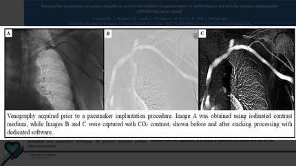
This single-center, prospective pilot study involved patients over 18 years referred for a CIED procedure. Patients with right-to-left shunts, pulmonary hypertension, respiratory failure, or known ICM allergies were excluded. Each patient underwent both ICM and CO2 venous angiography, with contrast administered through a catheter positioned in a forearm or antecubital vein. CO2 images were processed using digital subtraction angiography and stacking techniques with dedicated software (Figure 1). Two experienced radiologic technologists in electrophysiology imaging, blinded to the procedure and acquisition technique, evaluated the images. All patients provided written informed consent.
Of the sixty-two patients screened, two were excluded (one due to a patent foramen ovale with a right-to-left shunt and the other due to severe pulmonary hypertension). Among the 60 patients included, 46 (76.7%) were male, with a mean age of 72.6 ± 13.1 years, a mean creatinine level of 1.3 ± 0.7 mg/dL, and a mean estimated glomerular filtration rate (eGFR) of 61.2 ± 27.6 mL/min/1.73 m². Both ICM and CO2 angiography provided high-quality venous visualization, enabling the assessment of patency for the axillary and subclavian veins in over 90% of cases, with a slightly higher success rate for the axillary vein observed for ICM. ICM was more effective in visualizing the patency of the cephalic vein, while CO2 demonstrated an advantage in visualizing the patency of the jugular vein (Table 1). Four (6%) patients experienced CIN (defined as a 25% relative increase or a 0.5 mg/dL absolute increase in serum creatinine), while no allergic reactions were reported. No adverse events commonly associated with CO2 use in arterial applications, such as neurotoxicity, gas embolism, or pain, were observed in this study.
CO2 venography, utilizing digital subtraction and stacking technique, enables visualization of subclavian and axillary vein patency in over 90% of cases, making it a viable and safe option during CIED procedures. Further studies are warranted to confirm its clinical application, identify patient subgroups that may benefit from this approach, and assess its potential to reduce the risk of CIN.
Contributors

A Spadotto
Author
G Massaro
Author
M Carelli
Author
C Martignani
Author
M Ziacchi
Author
M Biffi
Author
I Diemberger
Author
You may be interested in