The relationship between left atrial electroanatomical voltage mapping and CMR-based fibrosis for enhanced preplanning in atrial fibrillation ablation
EP Europace Journal


Abstract
Cardiac magnetic resonance imaging-based late gadolinium enhancement (LGE-CMR) of left atrial (LA) fibrosis distribution can be valuable for preprocedural planning, patient selection and real-time guidance during atrial fibrillation (AF) ablation. However, the accuracy of LGE-CMR in assessing LA fibrosis is still debated.
To evaluate the accuracy of LGE-CMR-based LA fibrosis (CMR-fib) among ablation-naive patients compared to electroanatomical mapping (EAM-fib).
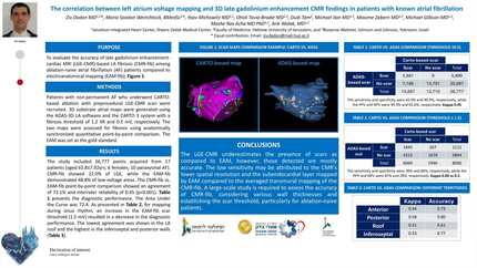
Patients with non-permanent AF who underwent CARTO-based mapping with preprocedural LGE-CMR scan were included. 3D substrate atrial maps were generated using the ADAS-3D-LA software and the CARTO 3 system with a fibrosis threshold of 1.2 and 0.5mV, respectively (Figure 1). The two maps were evaluated for fibrosis through anatomically synchronized, quantitative point-by-point comparison, with EAM used as the gold standard. Further analyses were performed based on the anatomical LA wall regions and, when available, CT-based LA wall thickness.
The study included 41,543 points acquired from 24 patients (aged 64±11 yrs; 7 females; 15 paroxysmal AF). CMR-fib showed 21% of LGE, while the EAM-fib demonstrated 42% of low-voltage areas. The CMR-fib vs. EAM-fib point-by-point comparison showed an agreement of 89% and interrater reliability of 0.53 (p<0.001). The sensitivity and specificity were 50% and 100%, respectively, while the PPV and NPV were 100% and 74%, respectively. The Area Under the Curve was 75. An increase in the EAM-fib fibrosis threshold (1.5mV) resulted in a decrease in the diagnostic performance.
The lowest agreement was observed in the LA roof, while the highest agreement was seen in the inferoseptal and anterior walls. Categorizing acquired points into different ranges of LA wall thickness (among 16 pts) revealed the highest agreement at diameters between 2.5 and 4 mm. In contrast, the agreement was lowest for diameters ≥4 mm and ≤0.5 mm (Figure 2).
LGE-CMR tends to underestimate the presence of fibrosis compared to EAM; however, the fibrosis that is detected is generally accurate. CT-based thickness measurements can be used for procedure preplanning to identify LA wall areas with enhanced CMR-based fibrosis accuracy. The low accuracy in mapping points at the outer ends of the scale suggests that the reduced sensitivity may be attributed to the lower spatial resolution of CMR, as well as the fact that EAM maps the subendocardial layer, while CMR-fib provides an averaged transmural mapping. A large-scale study is required to assess the accuracy of CMR-fib, considering various wall thicknesses and establishing the fibrosis threshold, particularly for ablation-naive patients.
Contributors

Z Dadon
Author
M Saadon Weinshtock
Author
M Shrem
Author
Y Michowitz
Author
O Tovia-Brodie
Author
D Tam
Author
M Ilan
Author
M Zabern
Author
M Glikson
Author
M Rav Acha
Author
A Wolak
Author
You may be interested in