Left atrial intramyocardial fat at pulmonary vein reconnection sites during atrial fibrillation redo ablation
EP Europace Journal


Abstract
Electrical reconnections between pulmonary veins (PVs) and the left atrium (LA) are frequently responsible for atrial fibrillation (AF) recurrences after pulmonary vein isolation (PVI)(1,2). Multidetector computed tomography (MDCT)-derived images can be post-processed to detect intramyocardial fat (inFAT) by signal density thresholding(3). InFAT might have a role for PV-LA reconnections.
To analyze the relationship between inFAT analyzed at pre-procedural MDCT-derived inFAT maps from first AF ablation and PV-LA reconnections in patients with AF recurrence undergoing redo ablation.
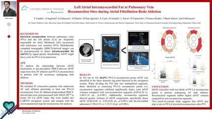
We included 45 consecutive patients who underwent AF redo ablation at our institution and had at least one PV-LA reconnection. First AF ablation preprocedural MDCT-derived data were post-processed to create 3D LA inFAT maps, that were loaded into navigation system for reconnection site analysis. 103 out of 156 (66.0%) PV-LA reconnection points colocalized with inFAT. Reconnection segments (PVI segments containing ≥1 PV-LA reconnection point) had higher volumes of total inFAT than non-reconnection segments (8.05±6.56 vs. 5.40±5.18 µL, p<0.001), as well as threshold subranges dense inFAT (0.06±0.06 vs. 0.03±0.04 µL, p<0.001) and fat-myocardial admixture (7.98±6.52 vs. 5.37±5.16 µL, p<0.001).
InFAT colocalize with two thirds of PV-LA reconnection points in patients undergoing AF redo ablation. Reconnection segments harbor higher inFAT volumes compared to non-reconnection segments. This proof-of-concept study suggests that inFAT may play a role in PV-LA electrical reconnections after PVI. Procedural workflow InFAT volumes dotplots
Contributors

F Landra
Author
A Saglietto
Author
G Falasconi
Author
D Penela
Author
D Soto-Iglesias
Author
E Curti
Author
B Tonello
Author
L Teresi
Author
D Turturiello
Author
P Franco-Ocana
Author
J Marti-Almor
Author
A Berruezo
Author
You may be interested in