Optimal image intensity ratio threshold for atrial focal monopolar pulsed field ablation lesions assessment by dark-blood late gadolinium enhancement cardiac magnetic resonance imaging
EP Europace Journal


Abstract
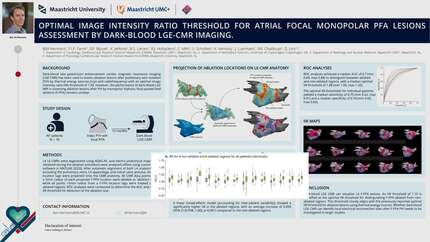
Dark-blood late gadolinium enhancement cardiac magnetic resonance imaging (LGE CMR) has been used to assess ablation lesions after pulmonary vein isolation (PVI) by thermal energy sources (cryo and radiofrequency) with an optimal image intensity ratio (IIR) threshold of 1.09. However, the performance of dark-blood LGE MRI in assessing ablation lesions after PVI by monopolar biphasic focal pulsed field ablation (F-PFA) remains unclear.
To determine the optimal IIR threshold for assessing F-PFA PVI lesions in dark-blood LGE MRI scans.
In eighteen patients, a 3D dark-blood LGE MRI was performed at 3 months after an initial PVI with F-PFA. Left atrial (LA) LGE MRIs were segmented using ADAS-AF, and electro-anatomical maps (EAM) obtained during the initial ablation procedure were analysed offline using custom-made software in MATLAB 2022b. After automatic alignment of both LA anatomies carefully excluding the pulmonary veins, LA appendage, and mitral valve annulus, the F-PFA location tags were projected onto the CMR anatomy. All CMR data points within a 5mm radius of each F-PFA location tags were treated as ‘ablation region’, while all points >5mm radius from a F-PFA location tags were treated as non-ablated regions. To maintain an equal ratio of ablated to non-ablated tissue, the same number of points as in the ablated region was randomly selected from the non-ablated region. Receiver operating characteristics (ROC) analyses were conducted to determine the area under the ROC curve (AUC) and the optimal IIR threshold for detection of the ablation scar. Average IIR values, AUC and optimal thresholds are determined for each patient. All values are reported are median and ranges [min-max] of these patient-specific values.
One patient was excluded from analysis due to a technical error during EAM export. The ablated region in the remaining seventeen patients consisted of a median of 3783 [1283-4568] IIR points. The median IIR of the ablated and non-ablated region was 1.14 [1.06-1.30] and 1.06 [0.98-1.10] respectively (Figure, top). A clear difference in IIR values between ablated and non-ablated tissue was seen for all but two patients. ROC analysis achieved a median AUC of 0.77 [0.44-0.88] to distinguish between F-PFA locations and non-ablated tissue (Figure, bottom left), with an average optimal IIR threshold of 1.10 [1.06-1.36].
Dark blood LGE MRI can visualize LA F-PFA lesions. An IIR threshold of 1.10 is identified as the optimal IIR threshold for distinguishing F-PFA locations from non-ablated tissue. This threshold closely aligns with the previously reported optimal IIR threshold for ablation lesions using thermal energy sources. Whether dark blood LGE MRI can identify local electrical reconnection sites after F-PFA PVI needs to be investigated in larger studies.
Contributors

B J M Hermans
Author
F I P Farnir
Author
G P Bijvoet
Author
K Jerltorp
Author
B S Larsen
Author
R J Holtackers
Author
C Mihl
Author
U Schotten
Author
K Vernooy
Author
J G L M Luermans
Author
S M Chaldoupi
Author
D Linz
Author
You may be interested in