Spatiotemporal dispersion and atrial fibrillation mapping: a novel stratifier of atrial cardiomyopathy?
EP Europace Journal


Abstract
Novel mapping techniques, such as machine learning algorithm-guided analysis of intracardiac electrograms for spatiotemporal dispersion (stDP), are promising tools for enhanced interventional substrate assessment. Recently, TAILORED-AF showed improved patient outcomes for atrial fibrillation (AF) ablation beyond pulmonary vein isolation. We investigated the relation of stDP to left atrial low-voltage regions.
This retrospective analysis included patients who underwent radiofrequency ablation with 3D electroanatomical mapping for voltage and activation in sinus rhythm and dispersion in AF at our clinic between May and October 2024. Patients with stDP were included and divided into two groups: one with stDP confined to low-voltage areas (LVA, <0.5 mV) and the other with stDP present in intermediate (0.5–1.5 mV) and normal voltage (>1.5 mV) regions. Primary demographic data and measurements of fibrotic left atrial cardiomyopathy were assessed for each group.
22 patients (7 female, 15 male) with a mean age of 66±10 years and an average body mass index (BMI) of 27.6±4.4 kg/m² met the inclusion criteria. AF had been diagnosed an average of 7±7 years before stDP mapping. First-time procedures were performed in six patients (27%). Four patients (18%) underwent more than two previous AF ablations.
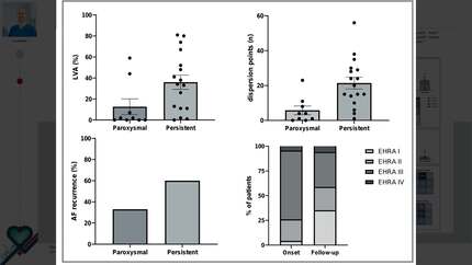
In 12 of 22 patients (55%, Group 1), stDP was detected exclusively within LVA, while in 10 patients (45.5%, Group 2), stDP was (also) registered in segments of intermediate and normal voltage. Both groups demonstrated no significant variation in age (G1: 67.8±8.7 vs. G2: 64±11.4, p=0.346), BMI (G1: 26.9±5.3 kg/m² vs. G2: 28.5±3.1 kg/m²; p=0.283), left ventricular ejection fraction (G1: 50±9 % vs. G2: 55±8 %; p=0.254), mean left atrial pressure (G1: 9.5±6.4 mmHg vs. G2: 7.6±2.8 mmHg; p=0.705), years from AF onset (G1: 6.25±4.6 vs. G2: 8.4±10; p=1) and number of previous AF ablations (G1: 1±0.9 vs. G2: 1.2±1; p=0.674). ProBNP levels showed a trend toward lower levels in the non-concurrent group (G1: 1513±1874 pg/ml vs. G2: 437±407 pg/ml; p=0.05). The left atrial volume index (LAVI) was significantly higher in Group 1 than in Group 2 (48.9±14.3 ml/m² vs. 33.8±1.3 ml/m²; p=0.011). Moreover, the LVA extent was substantially greater in the group with concurrent LVA and stDP detection (G1: 45.4±24.4 % vs. G2: 8.6±11.9 %; p<0.001).
Patients with more extensive LVA were more likely to show concomitant stDP. However, with a significantly smaller left atrial volume and lower proBNP levels but no difference in arrhythmia duration, the number of prior procedures, or left atrial mean pressure, our results suggest that spatiotemporal dispersion could be an early marker of atrial remodeling.
Spatiotemporal dispersion varies significantly between individual patients and might allow additional and early stratification of left atrial cardiomyopathy beyond the left atrial volume index and left atrial low voltage.
Contributors

E Heil
Author
J H Gerds-Li
Author
L H Boldt
Author
F Blaschke
Author
D Schoeppenthau
Author
A Parwani
Author
G Hindricks
Author
F Hohendanner
Author
You may be interested in