Efficiency and midterm outcomes of 3D imaging assistance in atrial fibrillation cryoablation
EP Europace Journal


Abstract
Evaluating the Efficiency, Safety, and Midterm Outcomes of 3D Imaging Assistance in Atrial Fibrillation Cryoballoon Ablation
Pulmonary vein (PV) isolation is a cornerstone therapy for symptomatic atrial fibrillation (AF). While 3D-navigation technologies commonly used with radiofrequency (RF) ablation significantly reduce X-ray exposure by providing enhanced anatomical detail, cryoballoon ablation (CBA) still relies on conventional fluoroscopy, resulting in considerable radiation exposure. Although modern fluoroscopy systems can integrate 3D imaging, the value of this capability for procedural guidance in CBA remains uncertain.
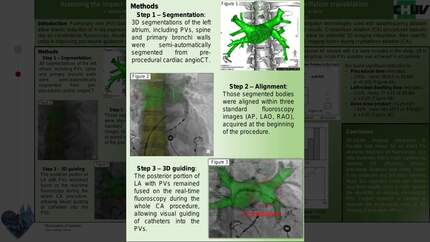
Pre-procedural cardiac CT was used to generate 3D segmentations of the left atrium, including PVs, spine, and primary bronchi, which were then fused with fluoroscopic images in three standard views (AP, LAO, RAO) at the procedure's start (Figure 1). We compared procedural characteristics and midterm outcomes between 3D-fusion-guided and conventional fluoroscopy-guided CBA. Efficacy was evaluated by assessing freedom from recurrence of sustained (>30 sec) atrial arrhythmia at 3, 6, and 12 months.
We included 44 patients (60±13 years, 85% male) with paroxysmal AF, split equally into 3D-guided and fluoro-only groups. Acute PV isolation was successful in all cases without major complications. The 3D-guided group had significantly shorter procedure times (81.5±14.8 vs. 95.6±16.6 min, p=0.004), reduced left-sided dwelling time (61.9±19.2 vs. 75.7±14.8 min, p=0.017), lower X-ray dose (Dose Area Product: 372.8±290 vs. 586.5±374 cGy*cm², p=0.045), and decreased fluoroscopy time (15.1±5.4 vs. 19.4±7.1 min, p=0.034). Additionally, 77% of 3D-guided procedures were completed in under 90 min, compared to only 32% in the control group (Figure 1). During the first year of follow-up, there was no significant difference in atrial arrhythmia recurrence between the groups (Figure 2).
3D-fusion imaging is feasible and provides precise PV anatomy on fluoroscopy, significantly enhancing CBA efficiency and safety without compromising efficacy in preventing atrial arrhythmia recurrence during a one-year follow-up period. Further research is warranted to confirm the long-term impact of 3D guidance on CBA outcomes. Fusion imaging for procedural guidance procedural time and follow up
Contributors

J Solana-Munoz
Author
C Teres
Author
C Ascione
Author
A Luca
Author
P Pascale
Author
G Domenichini
Author
E Pruvot
Author
M Le Bloa
Author
You may be interested in