RFA vs PFA in posterior wall isolation for PeAF: a single center prospective study - preliminary results
EP Europace Journal


Abstract
Despite advancements in mapping and ablation techniques, durable posterior wall isolation remains a challenge with conventional radiofrequency ablation, mainly due to anatomical difficulties in safely achieving transmural lesion due to the close proximity of the esophagus. Pulsed field ablation offers a promising new approach to ablation of the posterior wall, without the dangers to surrounding structures classically associated with RFA.
To compare the long term posterior wall isolation (PWI) durability with two ablation methods - RFA vs PFA.
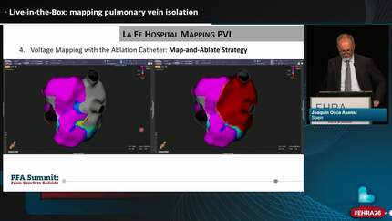
We performed a randomized trial including patients with PeAF assigned 1:1 to either PVI + PWI with RFA or to PVI + PWI with PFA. In the RFA group, PVI was achieved according to the CLOSE protocol and PEI was achieved with box-lesion set. In the PFA group, the PVI was performed with point-by-point PFA Cardiofocus Centauri system and PWI was achieved with contiguous area of ablations with interlesions distance of less than 6 mm. PVI and PWI was confirmed after a 20 minute waiting period with HD mapping. Follow-up procedure with HD re-mapping was performed 3 months after the index procedure to test durability of PVI and PWI.
There were 10 patients in the RFA group and 11 patients in the PFA. Baseline characteristics did not significantly differ between the 2 groups (male gender (90% vs 73%, p=0.15), mean age (62 ± 12,5 vs 59 ± 8,4 years, p=0.57), mean BMI (34,5 ± 10,5 vs 30,4 ± 4,6 kg/m2, p=0.40). Mean procedural duration was similar in RFA and PAF groups (212 ± 38,7 vs 198 ± 25,0 minutes, p=0.18). Both first pass PVI (RFA 7/10 vs PFA 11/11, p=0.03) and first pass PWI (RFA 4/10 vs PFA 11/11, p<0,05) during the index procedure differed significantly in both groups in favor of PFA. Follow-up reassessment was done in 7 patients in the RFA group and in 8 patients in the PFA group. Entrance block in all pulmonary veins per patient (5/7 vs 4/8, p=0.34) and per vein (25/28 vs 26/32, p = 0,20) did not significantly differ between the groups. In contrast, PWI was significantly higher in the PFA group compared with RFA group (⅞ vs 0/7, p<0,05).
PFA results in higher rate of PWI compared to RFA.
Contributors

You may be interested in