Impact of COngenital substrate 3d Reconstruction to guide vt Catheter Ablation. the CORECA study
EP Europace Journal


Abstract
In the presence of recurrent sustained monomorphic ventricular tachycardia (VT) catheter ablation may be an option in congenital heart disease (CHD). However, the unusual substrate and anatomy limit this option to few expert electrophysiologists.
We sought to investigate whether VT isthmus could be identified on 3D imaging substrate reconstruction from cardiac computed tomography scan (CT-scan) and/or magnetic resonance imaging (MRI) before ablation in CHD.
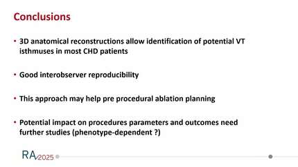
Forty consecutives patients with cardiac CT scan or MRI referred to VT ablation in 4 congenital electrophysiology centres were studied. Three electrophysiologists (EP) blinded from the ablation procedures and to each other's annotated ablation targets on 3d imaging substrate reconstruction (Figure 1). Once this was done, annotations were compared to each other and to ablation target on the electro-anatomical map performed during the procedure.
A total of 40 patients ( 27 male, 37 ±12 yo) with VT ablation in the setting of repaired tetralogy of Fallot (20), transposition of the great arteries (8), pulmonary atresia with VSD (3) or other CHD (9) were studied. In 5 patients, no VT isthmus was identified during the procedure but the substrate targeted was identified on imaging. In 30/40, the VT isthmus was identified on imaging. In the remaining 5, the VT isthmus was not identified on imaging. In one no scar was identified. In 2 patients, all 3 EP agreed on a strategy which did not include the isthmus and in 2 other patients, 2 EP agreed on a strategy not including the isthmus.
All 3 EP identified the same target in 28 patients, 2/3 EP in 10, 1/3 identify a target in 1 patient and no target was identified by any of the 3 EP in one patient.
3D substrate reconstruction identifies VT isthmus with certainty in 75% of patients referred for VT ablation in the setting of CHD. In 87.5%, it identifies substrate ablated during the procedure. rToF patient with cMRI Double Outlet RV pt with CT scan
Contributors

F Sacher
Author
F Bessiere
Author
V Waldman
Author
N Derval
Author
G Ditac
Author
P Pinon
Author
B Sacristan
Author
X Iriart
Author
S Bartoletti
Author
K Gardey
Author
L Iserin
Author
A Maltret
Author
H Cochet
Author
N Combes
Author
You may be interested in