Real-time MR-guided radiofrequency ablation and lesion evaluation in an MRI-compatible isolated beating pig heart platform
EP Europace Journal


Abstract
Cardiovascular magnetic resonance (CMR) is integral to the modern diagnosis and management of cardiac diseases, excelling in the precise depiction of cardiac anatomy, function, and tissue characterization. The evolution of interventional CMR (iCMR) as a real-time imaging technique for guiding intricate procedures such as myocardial ablation—without ionizing radiation—marks a significant advancement. However, progress is impeded by challenges in testing new MR-compatible instruments, validating MRI sequences, and correlating CMR data with histopathological findings, restricted by the limitations of in vivo tissue assessment. Our research uses a novel MRI-compatible, isolated beating pig heart platform to create a controlled environment for robust instrument testing and validation of advanced scanning protocols, bridging the gap between imaging characteristics and histological truth.
This proof-of-concept study evaluates the feasibility of performing real-time MR-guided radiofrequency ablation in an MRI-compatible isolated beating pig heart platform. The study also seeks to assess lesion characteristics through post-ablation MRI and histopathology.
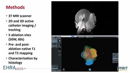
An isolated beating heart was harvested from a pig slaughtered for human consumption, prepared in compliance with regulatory guidelines, and connected to the custom-build MRI-compatible isolated pig heart platform. The platform supported left ventricular (LV) function in both Langendorff and working modes, using autologous heparinized blood at physiological temperatures and pressures. MR-guided catheter navigation and RFA were performed on a 3T MRI scanner with both 2D and 3D navigation using active catheter tracking. Pre- and post-ablation imaging included native T1 and T2 mapping to characterize tissue changes. Histological analysis was conducted after lesion creation.
Ablation was successfully performed with a power setting of 50 W for 60 seconds at five sites in the LV, leading to changes in both T1 and T2 relaxation time values in the ablated areas. Acute post-ablation imaging revealed an ablation core with significantly reduced T1 relaxation time values (≈1000ms), surrounded by areas of elevated T1 (≈1350ms) and T2 (≈80ms) values (reference T1: ≈1200ms, reference T2: ≈65ms), consistent with tissue necrosis and edema. Histological analysis confirmed a biphasic pattern of tissue injury, with a necrotic core and a surrounding rim of extravasation of erythrocytes.
This study demonstrates the feasibility of real-time MR-guided radiofrequency ablation in an MRI-compatible beating pig heart model, providing a controlled platform for refining MR-guided ablation techniques. The platform enables detailed MRI lesion visualization, histopathological correlation, and offers potential for advancing MR-guided therapies by improving lesion formation insights, testing equipment and ablation settings, and enhancing reproducibility in iCMR-guided procedures. Figure
Contributors

L H G Hopman
Author
J W M Niessen
Author
C P Allaart
Author
A J Nederveen
Author
E M Schrauben
Author
M J W Gotte
Author
J L Nelissen
Author
P J Reitzema
Author
R D Van Luijk
Author
M Koster
Author
B L M Smeets
Author
R W Boekhoven
Author
D Sunnarborg
Author
J Smink
Author
You may be interested in