Analysis of inflammatory and oxidative stress markers in radiofrequency and PFA for atrial fibrillation
EP Europace Journal


Abstract
Catheter ablation is a well-established treatment for atrial fibrillation and both radiofrequency (RFA) and pulsed field ablation (PFA) can be used as energy sources.Inizio moduloFine modulo
RFA uses thermal energy to induce tissue necrosis, while PFA relies on non-thermal, electric field-based electroporation to achieve similar results thus inducing cells apoptosis (1). These differences in energy delivery could influence the levels of inflammatory and oxidative stress markers during and after the procedure, which have been associated with AF recurrence rates.
Among the inflammatory markers, C-reactive protein (CRP) is often assessed to evaluate tissue response and systemic inflammation. Oxidative stress is another critical factor in AF ablation, with NOX2 (NADPH oxidase 2) serving as a key enzyme generating reactive oxygen species (ROS).
this prospective study included patients undergoing PVI for paroxysmal AF; the choice for the energy used for the ablation was left to the physician. Four blood samples were collected at different time points: the first from the femoral vein at the start of the procedure; the second was drawn from the left atrium immediately after the transseptal puncture; the third was obtained from the left atrium after having obtained the pulmonary veins isolation; and the fourth was collected by the anterocubital vein at 3 months since AF ablation. Radiofrequency Ablation was performed with Carto System and Smart Touch SF 4 mm catheter while PFA with Farawave Ablation Catheter.
a total of 63 patients were included in the study with comparable baseline characteristics, of which 32 underwent RF ablation, while 31 PFA ablation. Baseline CRP was 0.3±0.2 mg/ml for RF and 0.3±0.18 for PFA. Immediately after transeptal puncture CRP 0.3±0.3 was for RF (P>0.05) and 0.3±0.2 for PFA was (P>0.05). After the AF ablation CRP was 0.8±0.3 for RF (P Value<0.001) and for PFA was 0.4±0.24 (P>0.05). At 3 months follow-up CRP was 0.6±0.5 for RF (P<0.003) while it was 0.3±0.7 for PFA (P>0.05). Baseline NOX2 was 927±64 pg/ml for RF , and 918±83 pg/ml for PFA. Immediately after transeptal puncture NOX2 was 927±72 pg/ml for RF (P>0.05), and 918±7 pg/ml for PFA (P>0.05). After the AF ablation NOX2 was 2207±52 for RF (P<0.0001) and for PFA was 952±66 (P=0.07). At 3 months NOX2 was 1502±85 in RF (P<0.001) while it was 912±74 in PFA (P=0.8).
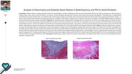
Radiofrequency ablation leads to an increase in inflammatory and oxidative stress markers in the left atrium following the ablation procedure compared to PFA. Characteristic lesion of PFA Characteristic lesion of RF
Contributors

N Pierucci
Author
M V M Mariani
Author
A P Piro
Author
D L Laviola
Author
V M L La Fazia
Author
M P Palombi
Author
S T Trivigno
Author
C L Lavalle
Author
You may be interested in