In-vivo endocardial and epicardial mapping of human sinus node: from electrical landmarks to anatomical landmarks
EP Europace Journal


Abstract
The human sinoatrial node (SAN) pacemaker is a complex structure located at the right atrium-superior vena cava (SVC) junction. It is intramural with a depth of 1 to 3 mm, and is composed of anastomosing clusters of specialized pacemaker myocytes. Recent clinical studies using simultaneous epicardial and endocardial mapping confirmed the existence of several discrete sinoatrial conduction pathways with distinct early activation sites or SAN exit zones (SAN-EZ).
This study aims to (1) perform in-vivo endocardial and epicardial electroanatomical mapping (EAM) of human SAN in inappropriate sinus node tachycardia (IST) and (2) correlate electrical findings with anatomical observations from thoracoscopy during hybrid SAN sparing IST ablation.
All consecutive patients with 1) Diagnosis of symptomatic IST, refractory or intolerant to drugs and 2) Endocardial and epicardial mapping of SAN during hybrid ablation were included. Local activation time (LAT) was defined using steepest -dV/dT on unipolar electrogram (EGM). SAN-EZ was defined as the earliest activation site on endocardial and epicardial maps. Endo-epicardial delay (EED) was the time difference between the first endo-epicardial activations. Endocardial–epicardial asynchrony (EEA) was defined as EED≥15 ms. Bipolar EGM morphology and SVC sleeves extension were analyzed. The presence of reversed polarity at SAN-EZ site was evaluated from the bipolar EGMs recorded from two contiguous electrode pairs of the mapping catheter. It was defined as a rapid simultaneous deflection in the opposite direction of the initial part of the bipolar EGMs occurring before the onset of the surface P-wave.
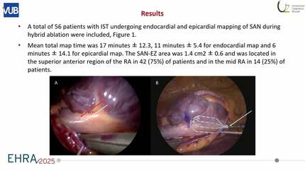
A total of 56 patients with IST undergoing endocardial and epicardial mapping of SAN during hybrid ablation were included, Figure 1. Mean total map time was 17 minutes ± 12.3, 11 minutes ± 5.4 for endocardial map and 6 minutes ± 14.1 for epicardial map. The SAN-EZ area was 1.4 cm2 ± 0.6 and was located in the superior anterior region of the RA in 42 (75%) of patients and in the mid RA in 14 (25%) of patients. The earliest activation occurred on epicardial SAN-EZ in all patients. The LAT of the epicardial vs endocardial SAN-EZ was -30.8 ms vs -12.4 ms, p<0.001. EED was 19.7 ms. EEA was present in 68% of patients. Unipolar EGM morphology at SAN-EZ sites was QS in all cases. Bipolar EGM reversed polarity at SAN-EZs was observed in 40 (71.4%) patients, Figure 2. SVC sleeves extension was 31.2 mm and inversely correlated with age.
Earliest SAN-EZ was found in the epicardium. Bipolar EGM reversed polarity is a novel electrophysiological marker for SAN-EZs. An increase in EED and EEA might be a marker of IST.
Contributors

C De Asmundis
Author
L Pannone
Author
I Eltsov
Author
D G Della Rocca
Author
A Sorgente
Author
I Overeinder
Author
G Bala
Author
A Almorad
Author
E Stroker
Author
J Sieira
Author
A Sarkozy
Author
P Brugada
Author
A Gharaviri
Author
G B Chierchia
Author
M La Meir
Author
You may be interested in