Validation of imageless electrocardiographic imaging for accessory pathway localisation in WPW syndrome
EP Europace Journal


Abstract
Catheter ablation is the definitive treatment for Wolff-Parkinson-White (WPW) syndrome, where precise preprocedural localisation of accessory pathways (APs) is critical to optimise outcomes and reduce procedural time. Traditional imaging and ECG techniques often fall short, particularly for septal APs. This study evaluated the diagnostic accuracy of an imageless electrocardiographic imaging (ECGi) system that does not require additional CT or MRI in localising APs for targeted ablation.
In this single-centre prospective study, adult and paediatric patients with WPW syndrome referred for AP ablation were consecutively enrolled. Non-invasive electroanatomic mapping was conducted using an imageless ECGi system, which uses a 128-electrode array to record body-surface potentials, a 3D torso model generated via a structured light camera, and an artificial intelligence algorithm to estimate the patient’s biventricular geometry. Epicardial electrograms were computed to obtain epicardial ventricular activation maps. The atrioventricular junction was divided into 11 regions using a modified Pappone classification to support AP localisation, with only the pathway responsible for ventricular pre-excitation being analysed in patients with multiple APs. The differential diagnostic capacity of the 12-lead ECG and ECGi was assessed by comparing the predicted AP locations with the ablation sites in invasive electroanatomical mapping (EAM) across three endpoints: (1) localisation within the same region, (2) within the same or adjacent regions, and (3) correct laterality (right, left, or septal).
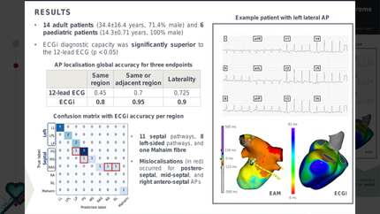
The study included 14 adult patients (mean age: 34.4 ± 16.4 years, 71.4% male) and six paediatric patients (mean age: 14.3 ± 0.71 years, 100% male). AP distribution included eleven septal pathways, eight left-sided pathways, and 1 Mahaim fibre. ECGi achieved a global accuracy rate of 80.0% in precisely localising APs to the correctly predefined AV region. When allowing for localisation in the adjacent area, the accuracy improved to 95.0%, and the accuracy in identifying laterality (right, left, or septal) was 90.0%. Notably, all mislocalizations were confined to septal pathways, suggesting the potential limitations of the current ECGi configuration in these complex anatomical regions. ECGi’s diagnostic capacity was significantly superior to the 12-lead ECG (p<0.05), which achieved an average accuracy of 45% for precise AP localisation, 70% when including the adjacent region, and 72.5% for laterality.
ECGi demonstrates a higher accuracy than the 12-lead ECG for AP localisation in WPW syndrome, supporting its use as a diagnostic tool for pre-ablation planning. Future integration of endocardial and epicardial mapping could improve accuracy for septal APs, further enhancing targeted ablation.
Contributors

S Monteiro
Author
J Reventos-Presmanes
Author
M Martinez-Perez
Author
G Portugal
Author
P S Cunha
Author
A M Climent
Author
M M Oliveira
Author
S M Laranjo
Author
You may be interested in