A concealed carcinoid cardiac metastasis uncovered by comprehensive cardiovascular magnetic resonance-based tissue characterization: a case report
European Heart Journal - Case Reports


Abstract
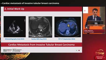
Cardiac metastases of carcinoid tumours are extremely rare, and their diagnosis poses a significant challenge. A variety of techniques has been reported in the literature for this purpose, ranging from echocardiogram to the Indium-111 Octreotide, positron emission tomography using specific tracers, and biopsy. Occasionally, the diagnosis is only made post-mortem. Recently, CMR (cardiovascular magnetic resonance) has been added to the diagnostic toolkit. This case report describes the CMR sequences that can be used to characterize cardiac metastases of carcinoid tumours.
A 55-year-old woman with an antecedent history of resected carcinoid tumour of the ileocecal junction underwent whole-body In-111 Octreoscan single-photon emission computed tomography in the context of her follow-up. This raised the suspicion of pericardial involvement, which prompted a CMR study. Comprehensive CMR findings were consistent with isolated carcinoid tumour metastasis embedded within the anterior papillary muscle. We describe the CMR sequences that were used to characterize the metastasis.
The rarity of cardiac metastasis of carcinoid tumour makes its diagnosis challenging and warrants a high level of clinical suspicion. Cardiovascular magnetic resonance imaging proves to be an indispensable tool in the tissue characterization of such tumours.
Contributors

Georgios Georgiopoulos
Author

Panagiota Mitropoulou
Author
Oxford University Hospitals NHS Foundation Trust Oxford , United Kingdom of Great Britain & Northern Ireland

Pier Giorgio Masci
Author
King's College London London , United Kingdom of Great Britain & Northern Ireland

Khan Tina
Author
Voges Inga
Author
Raisi Estabragh Zahra
Author
Minguito Carazo Carlos
Author
Thomson Ross
Author
You may be interested in