The role of transesophageal echocardiography in improving device size selection for percutaneous paravalvular leak closure
European Heart Journal - Cardiovascular Imaging


Abstract
Paravalvular leak (PVL) is an undesirable complication that can occur following valve implantations. The incidence of major leaks, which can clinically manifest as heart failure, hemolytic anemia, or both, has been reported to be 1-5% (1). The mortality rates for redo surgery are reported at 13%, 15%, and 37% for the first, second, and third operations, respectively (2). In recent years, percutaneous PVL closure (PPVLC) procedures, which are associated with lower mortality rates, have emerged as an attractive option for patients who are at high surgical risk.
Determining the appropriate device size for closing a defect is one of the most challenging aspects of PPVLC. Currently, there is no consensus on the optimal imaging modality for this purpose. We aimed to compare transthoracic and transesophageal echocardiographic measurements to accurately determine the device size.
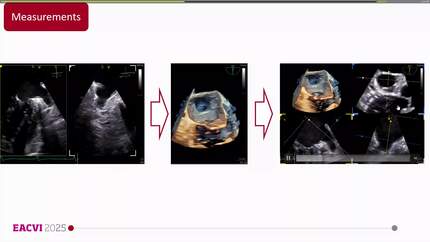
We reviewed our hospital records to identify patients diagnosed with moderate-severe and severe PVL from 2018 to 2024. A total of 81 who underwent percutaneous paravalvular leak closure (PPVLC) patients were evaluated. Eight of these 81 patients were excluded due to unsuccessful PPVLC, leaving 73 patients who were successfully treated with PPVLC were included. Technical success was defined as the deployment of the device without any interference with the prosthetic valve and with mild or less residual regurgitation. The defect size for all patients was evaluated using 2D transthoracic echocardiography (TTE), 2D transesophageal echocardiography (TEE), direct measurement of 3D TEE vena contracta, and 3D multi-planar reconstruction TEE (3D-MPR TEE).
A total of 73 patients with clinically significant moderate to severe or severe mitral and/or aortic paravalvular leak (PVL) were included in the study. Among these patients, 42 underwent aortic PPVLC and the remaining 31 underwent mitral PPVLC. Patients were categorized based on the implanted device size: 17 patients in the 6-7 mm group, 19 patients in the 8 mm group, 15 patients in the 10 mm group, 8 patients in the 12 mm group, and 14 patients in the 14-16 mm group. According to proportional odds logistic regression analysis, the 3D MPR TEE measurement was the strongest predictor of device size accuracy, both in the overall group and within the aortic/mitral subgroups. In the mitral subgroup, however, the predictive power of direct measurement of 3D TEE vena contracta and 3D MPR measurements was similar.
PPVLC has gained increasing interest, particularly over the last decade. Determining the appropriate device size is especially challenging in cases of mitral PVL. Our findings suggest that using 3D-MPR TEE measurements significantly improves the accuracy of device size selection in both mitral and aortic PVL. Additionally, direct measurement of 3D TEE vena contracta can serve as a viable alternative for patients with mitral PVL.
Direct measurement of 3D TEE
3D TEE MPR measurement
Contributors

M Tekin
Author
Mehmet Akif Ersoy Thoracic and Cardiovascular Surgery Training and Research Hospital Istanbul , Turkiye

G Babur Guler
Author
Mehmet Akif Ersoy Thoracic and Cardiovascular Surgery Training and Research Hospital Istanbul , Turkiye


A Guler
Author
Mehmet Akif Ersoy Thoracic and Cardiovascular Surgery Training and Research Hospital Istanbul , Turkiye
K Memic Sancar
Author
Mehmet Akif Ersoy Thoracic and Cardiovascular Surgery Training and Research Hospital Istanbul , Turkiye
M Cicek
Author
Mehmet Akif Ersoy Thoracic and Cardiovascular Surgery Training and Research Hospital Istanbul , Turkiye
You may be interested in