Sizing of the aortic valve using virtual reality - next step towards personalized medicine?
European Heart Journal


Abstract
High-quality imaging is integral throughout the transcatheter aortic valve implantation (TAVI) process, from patient selection and preprocedural planning to real-time guidance during the procedure and postprocedural follow-up. Multislice computed tomography (MSCT) is currently the gold standard. The next step in this area is the three-dimensional (3D) visualization of cardiac and extra-cardiac structures using virtual reality (VR).
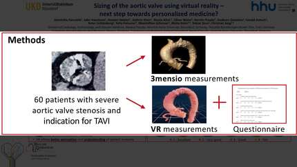
This study aimed to investigate the feasibility, accuracy, and reproducibility of VR for the visualization of the aortic valve, the surrounding structures, and its role in the planning of TAVI procedures.
Based on the preprocedural MSCT data of 60 patients who underwent TAVI at our heart center, measurements using 3mensio software were compared with 3D visualizations and measurements using VR software. Additionally, eight physicians evaluated the application of VR using a structured questionnaire to define its benefits and limitations.
VR measurements correlated well with the 3mensio software measurements, and there were no significant differences between the two methods. The VR measurements were mentioned first: Annulus perimeter 77.4 ± 7.8 vs. 77.0 ± 8.1 (p=0.182), r = 0.929; Annulus diameter 24.6 ± 3.2 vs. 24.4 ± 2.6 (p=0.483), r = 0.868; LVOT diameter 23.5 ± 3.1 vs. 23.7 ± 2.8 (p=0.442), r = 0.785; SoV left 32.9 ± 4.1 vs. 32.7 ± 3.7 (p=0.331), r = 0.920; SoV right 30.8 ± 4.3 vs. 30.7 ± 3.7 (p=0.653), r = 0.909; noncoronary SoV 32.9 ± 3.6 vs. 32.6 ± 3.7 (p=0.209), r = 0.913; STJ 30.0 ± 3.6 vs. 29.7 ± 3.2 (p=0.175), rs = 0.882; Ascending aorta 34.5 ± 3.4 vs. 34.5 ± 3.1 (p=0.469), r = 0.921, RCA height 15.1 ± 3.2 vs. 15.4 ± 3.2 (p=0.179), rs = 0.910, LCA height 12.4 ± 2.7 vs. 12.3 ± 3.0 (p=0.252), rs = 0.924. Furthermore, users reported that VR simplified anatomical understanding (1.3 ± 0.5 vs. 1.9 ± 0.4, p = 0.046), and improved 3D comprehension (1.1 ± 0.4 vs. 2.3 ± 0.5, p = 0.023).
The use of VR in the planning of TAVI procedures enables precise and reproducible measurements. In addition, VR can help assess the often complex anatomy, which can increase the safety and efficiency of procedures. Further clinical studies are needed to evaluate the potential benefits of VR application in planning and performing other structural interventions.
Contributors

J Haschemi
Author
S Afzal
Author

K Piayda
Author
G Antoch
Author
F Fleissner
Author
M Scherner
Author
M Kelm
Author

You may be interested in