Late iodine enhancement quantification by computed tomography: a comparison study against cardiac magnetic resonance imaging
European Heart Journal


Abstract
Cardiac computed tomography (CT) has emerged as a promising alternative to cardiac magnetic resonance (CMR) for myocardial characterization. However, the quantification of late iodine enhancement (LIE) and its threshold values have not been previously reported. The Full Width Half Maximum (FWHM) technique established a cut-off value of 50% of the maximum intensity signal to quantify late gadolinium enhancement (LGE) in CMR. Nevertheless, the distinction between healthy and fibrotic myocardium in CT is less pronounced, prompting the need for specific threshold values for accurate quantification of LIE with this technique.
Our primary aim was to validate and determine the optimal cut-off values for quantifying LIE using cardiac CT.
We included consecutive patients who underwent both LIE and LGE within a period of less than 2 months. Late enhancement quantification was performed using a commercially available software (figure 1). The FWHM technique was applied for LGE quantification. Various signal density cut-off values (70%, 75%, 80%, 85%, and 90%) were explored for LIE quantification. The total myocardial mass, total scar mass and scar proportion were calculated. The Intraclass Correlation Coefficient (ICC) was computed for each of the specified cut-off values.
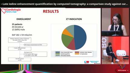
From November 2022 to February 2024, 58 patients underwent cardiac CT with LIE. Among them, 17 patients (48 (33;59) years; 13 (68,4%) male) had concomitant LGE and were included in the final analysis. The most common indication for CT was myocardial infarction with non-obstructive coronary arteries (52,9%).
The total myocardial mass by CT was 108.83 ± 21.69 g, while the total myocardial mass by CMR was 100.33 ± 25.19 g. The ICC between both techniques was good (0.78, p=0.001).
A cut-off value of 75% for the CT showed the strongest correlation with the CMR, both for total scar mass and scar proportion. Total scar mass was 9.43 ± 8.29 g for CT and 10.7 ± 8.46 g for CMR, with an ICC of 0.68 (p=0.01). Scar proportion was 9.97 ± 8.26% for CT and 11.09 ± 9% for CMR, showing a good correlation (ICC 0.73, p=0.006).
Late iodine enhancement quantification by cardiac computed tomography is feasible. A signal density threshold of 75% showed the strongest correlation with the cardiac magnetic resonance. Example of late iodine enhancement
Contributors


You may be interested in