3D modelling-based left atrium geometry analysis to predict atrial fibrillation in patients with cryptogenic stroke
European Heart Journal


Abstract
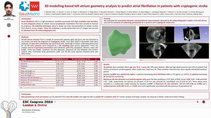
Atrial fibrillation (AF) is a high prevalence condition associated with high morbidity and mortality. Its early diagnosis allows to initiate several prophylactic treatments that have proved to improve its prognosis. 3D modeling techniques allow to design accurate representations of cardiac cavities from diverse source images.
We evaluated the association between new geometrical measurements obtained by 3D echocardiographic models of the left atrium (LA) and the presence of underlying paroxysmal AF in patients with cryptogenic stroke (CS).
3D LA volumes from a sample of consecutive patients aged ≥60 years old and admitted to our Stroke Unit with the diagnosis of CS from April 2019 to November 2020 were analyzed. 15 days ECG monitoring was performed after discharge to detect underlying paroxysmal AF. All 3D echo volumes were analyzed by a 3D modeling open-source application. From this analysis several LA geometry parameters were obtained: sphericity, elongation, flatness, major axis length, minor axis length, maximum 2D diameter (max2Ddiam) and surface-volume ratio (S/V). Functional atrial parameters (3D left atrial ejection fraction, LAEF) were also determined.
36 patients were analyzed. Mean age was 78 ± 7 years old. 19% were diabetic, 58% had high blood pressure and 14% of patients had history of ischemic cardiomyopathy. Mean body-mass index was 26. These baseline characteristics were equally distributed between groups.
Baseline proBNP was significantly higher in patients developing atrial fibrillation (949 vs 275 pg/mL; p= 0,014.).
Maximum 2D diameter (max2Ddiam) and Surface-volume ratio (S/V) were the best predictors of AF (AUC of ROC curves: 0.69; 0.49 – 0.88 and 0.63; 0.42 – 0.84, respectively). An optimal cut-off point of 39 mm was selected for max2Ddiam and of 0.20 for S/V. These values were included in a multivariate model adjusted by baseline proBNP. A Max2Ddiam <39 mm and a S/V ≥0.20 were both significantly associated with the presence of paroxysmal AF (OR 6.96; p= 0.048 and OR 12.22; p = 0.043, respectively).
New geometrical atrial parameters can be obtained from echo 3D models and might be able to predict AF in patients with CS. Further analysis and larger samples are required to better understand these findings. Table Figure
Contributors



D Viliani
Author
S Montejano
Author
G Diego Nieto
Author
A Benedicto Buendia
Author
L Vilches Miguel
Author
B Santos Martin
Author
B Lopez Melgar
Author
L Dominguez Arganda
Author

A Torrado Carvajal
Author
L J Jimenez-Borreguero
Author
You may be interested in