Case report: a non-invasive approach to diagnosis and management of pericardial haemangioma
European Heart Journal - Case Reports


Abstract
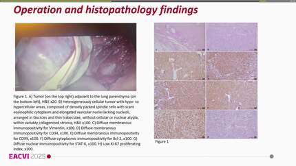
Pericardial haemangiomas represent a very rare subset of benign cardiac tumour in an unusual location, posing a diagnostic and clinical challenge. Historically, the definitive diagnosis was achieved through surgical resection or at biopsy. In recent years, multi-parametric cardiac magnetic resonance imaging (MRI) has proven to offer a non-invasive, biopsy-like approach to tumour characterization.
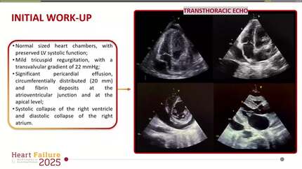
In our case, multimodality imaging was used to characterize a pericardial mass as a haemangioma discovered coincidentally with a brain glioma. Diagnostic certainty was substantially improved through utilization of successive post-contrast bright-blood imaging at cardiac MRI, demonstrating a characteristic enhancement pattern of haemangiomas in direct comparison to the blood pool. Conservative management and mid-term follow-up showed an uneventful clinical course and partial regression of the presumed pericardial haemangioma.
In the presence of typical features and application of individually tailored protocols, multimodality imaging can characterize cardiac tumours and guide patient management so that more invasive measures may be avoided. In our case of a suspected pericardial haemangioma, a conservative strategy was adopted with clinically uneventful course over a 2-year period. Whether this strategy can be applied to other patients with this rare tumour remains unclear, but the case report provides important information about the natural history of this entity and tissue characterization by cardiac MRI.
Contributors

Dominik A Steffen
Author
Laurene Suter-Magpantay
Author
André Linka
Author
Lucas Weber
Author
Sarandeep Kaur Marwaha
Author
Ahsan Aftab Khan
Author
Sumit Sohal
Author
Wissam Alajaji
Author
Deborah Cosmi
Author
Nicolo Sisti
Author
You may be interested in