Value of high-output pace-mapping of the right phrenic nerve for enabling safe radiofrequency ablation of atrial fibrillation: insights from three-dimensional computed tomography segmentation
EP Europace Journal


Abstract
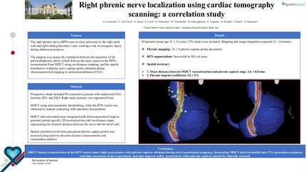
Right phrenic nerve (RPN) injury is a disabling but uncommon complication of atrial fibrillation (AF) radiofrequency ablation. Pace-mapping is widely used to infer RPN’s course, for limiting the risk of palsy by avoiding ablation at capture sites. However, information is lacking regarding the distance between the endocardial sites of capture and the actual anatomic RPN location. We aimed at determining the distance between endocardial sites of capture and anatomic CT location of the RPN, depending on the capture threshold.
In consecutive patients undergoing AF radiofrequency ablation, we defined the course of the RPN on the electroanatomical map with high-output pacing at up to 50 mA/2 ms, and assessed RPN capture threshold (RPN-t). The true anatomic course of the RPN was delineated and segmented using CT scan, then merged with the electroanatomical map. The distance between pacing sites and the RPN was assessed. In 45 patients, 1033 pacing sites were analysed. Distances from pacing sites to RPN ranged from 7.5 ± 3.0 mm (min 1) when RPN-t was ≤10 mA to 19.2 ± 6.5 mm (min 9.4) in cases of non-capture at 50 mA. A distance to the phrenic nerve > 10 mm was predicted by RPN-t with a ROC curve area of 0.846 [0.821–0.870] (
These data emphasize the utility of high-output pace-mapping of the RPN. Non-capture at 50 mA/2 ms demonstrated very high specificity for predicting a distance to the RPN > 10 mm, ensuring safe radiofrequency delivery.
Contributors

Gregory Supple
Author
Ioan Liuba
Author
Michal Wasiak
Author
Erica Zado
Author
Benoit Desjardins
Author

You may be interested in