Calcium density of the aortic valve: a comparison of its measurement between echocardiography and computed tomography in a cohort of South American patients with severe aortic stenosis
European Heart Journal - Cardiovascular Imaging


Abstract
The baseline calcium load of the valve is indicative of aortic stenosis progression. While echocardiography remains the standard study for severity assessment, with acknowledged limitations, computed tomography excels in its remarkable reproducibility in measurements, particularly through the calculation of the valve calcium score. The indexing of valve calcium to the area of the aortic ring, termed the calcium density of the aortic valve, has been proposed as an additional diagnostic and prognostic variable. In Asian populations, the diagnostic precision of the calcium score threshold is lower, suggesting that valve area density might be a more suitable reference. Local-level research on this variable is warranted.
Correlate the measurement of aortic valve area density performed by computed tomography with that calculated using echocardiographic variables. Analyse whether the density of the aortic valve area has prognostic value concerning the presence of paravalvular leak one month after transcatheter aortic valve implantation (TAVI).
Analytical retrospective cohort study with prospective follow-up aimed at correlating the measurement of aortic valve area density performed by computed tomography with that calculated using echocardiographic variables and assessing its prognostic value. The registry was conducted at two centres. All patients with symptomatic severe aortic stenosis who underwent TAVI from June 2021 to June 2023 were included. The ethical guidelines were followed, respecting the anonymity of the data and the Declaration of Helsinki. Statistical analyses were performed using SPSS V.25, with a P value less than 0.05 considered significant.
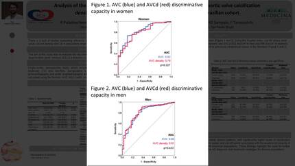
Sixty-three patients were included, 47.61% of whom were female, with a mean age of 82 years (Q1-Q3: 79; 86 years). A statistically significant correlation was found when assessing the density of the aortic ring area estimated from echocardiography compared to that estimated from computed tomography (Figure 1). In the examination of the ROC curves for the calcium score and the aortic valve calcium density, they were identified as poor predictors for paravalvular leak outcomes (Figure 2).
A correlation is observed between the density of the aortic valve area measured by computed tomography and that calculated with echocardiographic variables. However, they do not appear to hold prognostic value regarding the presence of a paravalvular leak one month after the TAVI procedure.
Contributors


You may be interested in