Imaging-derived tissue visualization guides catheter ablation of premature ventricular contractions
EP Europace Journal


Abstract
The presence of myocardial scarring is associated with worse outcomes after catheter ablation of premature ventricular contractions (PVCs). Three-dimensional substrate characterization is emerging for peri-procedural ablation guidance, but its value for tissue visualization during PVC ablation is unknown.
The aim of the present study was to assess the impact of imaging-derived tissue visualization on lesion planning, procedural and clinical outcomes of PVC ablation.
Thirteen consecutive patients (92.3% male, 62.2±12.4 years) undergoing unipolar (n=8) or bipolar (n=5) PVC catheter ablation with integrated three-dimensional imaging from multidetector computed tomography or cardiac magnetic resonance imaging were included. All patients presented with assumed myocardial scarring due to an underlying cardiomyopathy and/or after previous ablation.
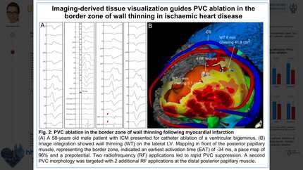
Among all patients (30.8% (peri-)myocarditis, 20.0% ischemic, 15.4% dilated cardiomyopathy, 15.4% idiopathic, 7.7% nonischemic, 7.7% congenital heart disease) undergoing ablation of 20 PVC morphologies (median 1 (IQR 1-2) per patient; 75.0% non-outflow tract; 70.0% left-sided), myocardial scarring was present in 11/13 (84.6%). Wall thinning <5 mm covering an area of 36.1±17.8 cm² contained the PVC origin in 4/13 (30.8%) patients and was safely targeted with radiofrequency lesions with up to 45 W (maximum power: median 32 W (IQR 30-35)) (Figure 1). Imaging-guided bipolar ablation was performed in 5/8 (62.5%) patients with previously therapy-refractory PVCs including foci at the left ventricular summit and the right ventricular outflow tract each in 2 and the left coronary cusp in one patient (Figure 2). PVCs were efficiently suppressed with less radiofrequency lesions (5.4±3.8 vs. 55.8±36.9; p=0.0160) with shorter duration (4.1±3.2 min vs. 23.3±13.5 min; p=0.0152) compared to prior unipolar ablation. Bipolar lesions resulted in a higher impedance (158 Ω (IQR 101-174) vs. 106 Ω (IQR 99-124); p=0.0025), while requiring a lower temperature (36 °C (IQR 35-38) vs. 39 °C (IQR 36-43); p=0.0001) and less power (26 W (IQR 20-30) vs. 30 W (IQR 25-31); p=0.0376). Complete suppression of the clinical PVC was reached in all 13 patients. During a follow-up of 396.8±299.7 days, PVC burden decreased from 20.1±14.3% to 1.7±1.6% (p=0.0172).
Imaging-derived tissue visualization enables determination of myocardial wall thinning <5 mm as a substrate for recurrent PVCs, which can be safely targeted. Three-dimensional imaging efficiently guides high power lesion application and bipolar ablation of PVCs at anatomically challenging foci. Anatomically challenging foci Imaging-guided bipolar ablation
Contributors


You may be interested in