Atrial fibrillation cryoablation guided by pulmonary vein pressure: comparison with the standard strategy
EP Europace Journal


Abstract
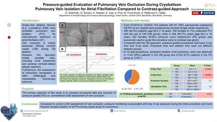
Pulmonary vein (PV) isolation is the cornerstone for the treatment of patients with atrial fibrillation (AF). Balloon cryoablation (BCA) is the preferred single-shot option for ablation of PVs using contrast guided occlusion as the standard approach (SA). We have made a simplified procedure, using a pressure-guided approach (PG) aimed to obtain a PV wedge curve. This technique has been described previously(1-3). To our knowledge, no comparison has been made in real life with a mixed patient setting in AF (paroxysmal and persistent) and non-selected anatomy with exclusive PG.
To compare a prospective cohort of patients going into AF BCA, using SA versus PG.
Between January 2022 and October 2023, 50 consecutive patients underwent BCA. The first 25 patients were treated with SA protocol and the latter 25 with PG. In both techniques, the aim was to obtain PV isolation with a 240 seconds application and no bonus attempt. A computed tomography angiography or MRI of the left atrium (LA) was performed in all the patients. 28-mm Cryoballoon and Intracardiac echo were used in all the procedures. We compared patients and procedure characteristics between both techniques.
Both groups (SA vs PG) showed similar baseline characteristics with not significant differences between them: mean age (58±10 vs 61±10 years), median LA diameter (40±8 vs 41±5mm), mean LVEF (60±9 VS 63±7%), Paroxysmal AF (60 vs 56%), average CHA2DS2-VASc score (1.3±1.3 vs 1.8±1.5), One patient is SA group had a previous radiofrequency (RF) ablation with 4/4 veins reconnected, and in the PG group two patients had RF ablation with 7/8 veins reconnected (Table 1). The procedural characteristics between SA and PG, also did not show significant differences: median procedural time (120±20 vs 130±38 min), fluoroscopic time (24±13 vs 30±19 min), and isolation was achieved in 98/99 veins (99%) in each group (Table 2). In the SA group, two patients had complications: one patient with a transient palsy of the phrenic nerve and because of this, only 3/4 veins were targeted, and the second patient had a post-procedure pericarditis. In the PG group, one patient had a pericardial effusion seen at the end of the procedure that did not require drainage.
In our initial experience, BCA for AF with both strategies proved to be safe and effective in this mixed group with non-selected anatomy. This result reassures the PG as a way to enhance CBA because of the advantage in avoiding the use of intraprocedural IV contrast. Impact in performance during follow-up is yet to be seen in our cohort. Baseline characteristics Procedural characteristics
Contributors

You may be interested in