Giant left sinus of Valsalva aneurysm as a rare cause of acute myocardial infarction: a case report
European Heart Journal - Case Reports


Abstract
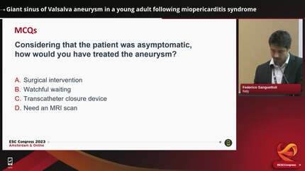
Sinus of Valsalva aneurysm (SVA) is a rare but potentially life-threatening condition. Acute myocardial infarction (MI) is a rare consequence of aneurysmal dilatation of one or more sinuses of Valsalva. We present a case of an unruptured and partially thrombosed left SVA, presenting as anterior MI and congestive heart failure.
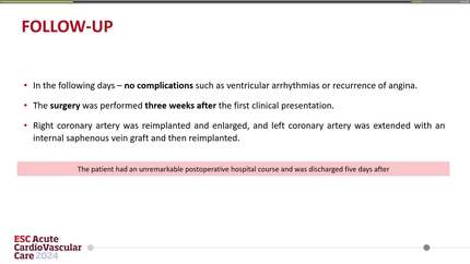
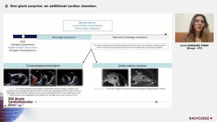
A 55-year-old gentleman was admitted with pulmonary oedema and a late presenting ST-elevation MI with Q wave. After initial treatment on furosemide infusion, a coronary angiography showed significant stenosis in both his left main stem (LMS) and left anterior descending artery (LAD). This is likely a result of external compression, potentially from the enlarged left sinus of Valsalva. A subsequent transthoracic echocardiogram and transoesophageal echocardiogram (TOE) confirmed large SVA involving the left coronary cusp measured 9.9 cm compressing both LMS and LAD.
Left SVAs are rare and frequently asymptomatic, typically being identified incidentally. Due to the close proximity of the left coronary system, they can present with myocardial ischaemia due to extrinsic compression of the coronary system. We were able to perform a comprehensive multi-modality assessment of left SVA, which helped establish this unusual diagnosis and guide management. Transthoracic echocardiogram and TOE helped assess the SVA and demonstrated the thrombus
Contributors


Mengshi Yuan
Author
William Moody
Author
Shanat Baig
Author
Sohail Q Khan
Author
Dimitrios A Vrachatis
Author
A Shaheer Ahmed
Author
Parag Bawaskar
Author
Ralph Mark Louis Neijenhuis
Author
You may be interested in