Hibernoma: a case report of a rare cardiac tumour
European Heart Journal - Case Reports


Abstract
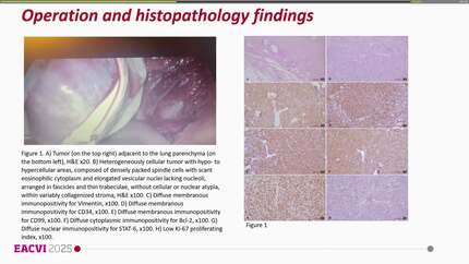
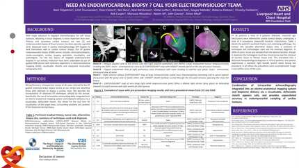
A cardiac hibernoma is a rare phenomenon, with just a handful of reports in the literature. They are difficult to characterize with conventional imaging including echocardiography, computed tomography (CT), cardiac magnetic resonance (CMR), or positron emission tomography (PET). Their definitive diagnosis relies primarily on histopathology via either endovascular or surgical biopsy. Previous case reports have entailed surgical excision followed by histopathology; however, surgery may be unfavourable in some patients with increased perioperative risk.
We present the case of a 57-year-old woman who was referred to our cardiology service with an interatrial lipomatous mass found incidentally on chest CT for assessment of rib fractures. She had 6 months of unexplained syncope, which was attributed to superior vena cava (SVC) compression demonstrated by chest CT. The mass had benign characteristics on echocardiography, CT, and CMR but was glucose-avid on PET, which indicated a possible malignancy such as liposarcoma. Her comorbid and very significant airways disease precluded her from surgical excision, so instead, endovascular biopsy was performed. Histopathology showed brown fat which was negative for mouse double minute 2 amplification on fluorescence
The definitive diagnosis of a cardiac hibernoma is complex and relies heavily on histopathology due to the contradictory findings on chest imaging. Careful consideration of management within a multidisciplinary team setting is essential to achieve a successful outcome.
Contributors

Farrah Othman
Author
Rooshdiya Karim
Author
Rajesh Puranik
Author
Christoph Jensen
Author
Michael Z L Zhu
Author
Subhi Akleh
Author
Nikesh Jathanna
Author
You may be interested in