Added-value of molecular imaging in myocardial metastasis of an ileal neuroendocrine tumour treated with peptide receptor radionuclide therapy: a case report
European Heart Journal - Case Reports


Abstract
Neuroendocrine tumours (NET) conform a rare type of neoplasm, mostly located in the gastrointestinal tract. They are slow-growing tumours, so at the time of the diagnosis, most patients present with metastatic lesions, mainly in the liver. The myocardium is a rare and important organ for metastasis, in which 68Ga-Dotatate positron emission tomography-computed tomography (PET/CT) shows a high diagnostic sensitivity for its detection, contrary to carcinoid valve disease, where anatomic imaging plays a key role, especially the echocardiogram.
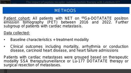
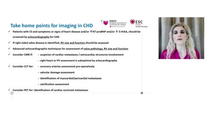
A 60-year-old man diagnosed with metastatic progressive ileal NET, who underwent a 68Ga-Dotatate PET/CT prior 177Lu-Dotatate therapy, showed a metastatic lesion in the left ventricle that was undetected in previous studies, such as an Octreoscan® and CT. A transthoracic echocardiogram was performed revealing the existence of a second cardiac lesion, a tricuspid valve carcinoid disease. A cardiac magnetic resonance showed no late gadolinium enhancement.
The 68Ga-Dotatate PET/CT is currently considered the gold standard for assessment and follow-up of NET, including those with rare sites of metastasis such as cardiac infiltration. In this case, it stimulated the persue of possible cardiac involvement, detecting the coexistence of two types of lesions (cardiac metastasis and carcinoid valve disease). Of these, carcinoid valvulopathy develops in 50% of NET cases, while cardiac metastasis (CM) is less frequent (only 5%).
Contributors

Estephany Abou Jokh Casas
Author
University Hospital of Santiago de Compostela Santiago de Compostela , Spain
Charigan Abou Jokh Casas
Author
Virginia Pubul Núñez
Author
Tina Khan
Author
Joseph Moutiris
Author
Zahra Raisi Estabragh
Author
Richard Alexander Brown
Author
Edwina McNaughton
Author
Ross Thomson
Author
You may be interested in