Case report: intravenous leiomyomatosis with intracaval and intracardiac growth
European Heart Journal - Case Reports


Abstract
Intravenous leiomyomatosis (IVL) is a rare, benign smooth muscle cell tumour that extends beyond the pelvis. These tumours grow within vascular channels and can progress to involve the heart and pulmonary vasculature.
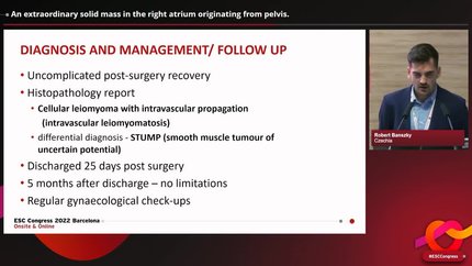
A 44-year-old female initially presented to her primary care physician for subacute bloating. In the weeks leading up to her presentation, she was in good health. On admission, computed tomography (CT) imaging of the abdomen and pelvis was notable for a mixed solid and cystic mass arising from the fundal myometrium with invasion into the inferior vena cava (IVC). Transthoracic echocardiogram (TTE) was notable for mobile mass in the right atrium originating from the IVC. The mass was further evaluated by cardiac magnetic resonance (CMR) imaging before a multidisciplinary, single-staged thoracoabdominal resection was performed. The procedure was well tolerated, and the entire mass was successfully removed without complication. Subsequently, pathological analysis of the resected tumour revealed benign smooth muscle cells, confirming the diagnosis of IVL.
Intravenous leiomyomatosis is a rare cause of right-sided cardiac tumours but should be considered in premenopausal females, even those with a prior history of hysterectomy. The clinical presentation of patients with IVL is varied and imaging including CMR, CT, and TTE to evaluate the tissue characteristics and source of the cardiac mass should be performed. Finally, while imaging revealing a freely mobile pelvic mass extending into the IVC and right heart chambers is strongly suggestive of IVL, definitive diagnosis requires pathological analysis of resected tissue.
Contributors

Daniel S Kikuchi
Author
Clive A Goulbourne
Author
Kristen D Starbuck
Author
Marcelo F Fernandes
Author
Konstantinos Stathogiannis
Author
Domenico Filomena
Author
Nikesh Jathanna
Author
Sara Monosilio
Author
You may be interested in